Neurosteroid transport by the organic solute transporter OSTα-OSTβ
- PMID: 20649839
- PMCID: PMC2939961
- DOI: 10.1111/j.1471-4159.2010.06920.x
Neurosteroid transport by the organic solute transporter OSTα-OSTβ
Abstract
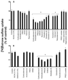
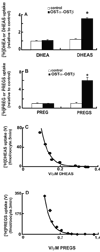
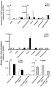
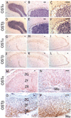
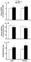
A variety of steroids, including pregnenolone sulfate (PREGS) and dehydroepiandrosterone sulfate (DHEAS) are synthesized by specific brain cells, and are then delivered to their target sites, where they exert potent effects on neuronal excitability. The present results demonstrate that [(3)H]DHEAS and [(3)H]PREGS are relatively high affinity substrates for the organic solute transporter, OSTα-OSTβ, and that the two proteins that constitute this transporter are selectively localized to steroidogenic cells in the cerebellum and hippocampus, namely the Purkinje cells and cells in the cornu ammonis region in both mouse and human brain. Analysis of Ostα and Ostβ mRNA levels in mouse Purkinje and hippocampal cells isolated via laser capture microdissection supported these findings. In addition, Ostα-deficient mice exhibited changes in serum DHEA and DHEAS levels, and in tissue distribution of administered [(3)H]DHEAS. OSTα and OSTβ proteins were also localized to the zona reticularis of human adrenal gland, the major region for DHEAS production in the periphery. These results demonstrate that OSTα-OSTβ is localized to steroidogenic cells of the brain and adrenal gland, and that it modulates DHEA/DHEAS homeostasis, suggesting that it may contribute to neurosteroid action.
© 2010 The Authors. Journal Compilation © 2010 International Society for Neurochemistry.
Conflict of interest statement
The authors affirm that there are no conflicts of interest.
Figures



Similar articles
-
Neurosteroid Transport in the Brain: Role of ABC and SLC Transporters.Front Pharmacol. 2018 Apr 11;9:354. doi: 10.3389/fphar.2018.00354. eCollection 2018. Front Pharmacol. 2018. PMID: 29695968 Free PMC article. Review.
-
Functional complementation between a novel mammalian polygenic transport complex and an evolutionarily ancient organic solute transporter, OSTalpha-OSTbeta.J Biol Chem. 2003 Jul 25;278(30):27473-82. doi: 10.1074/jbc.M301106200. Epub 2003 Apr 28. J Biol Chem. 2003. PMID: 12719432
-
Heterodimerization, trafficking and membrane topology of the two proteins, Ost alpha and Ost beta, that constitute the organic solute and steroid transporter.Biochem J. 2007 Nov 1;407(3):363-72. doi: 10.1042/BJ20070716. Biochem J. 2007. PMID: 17650074 Free PMC article.
-
OSTalpha-OSTbeta: a major basolateral bile acid and steroid transporter in human intestinal, renal, and biliary epithelia.Hepatology. 2005 Dec;42(6):1270-9. doi: 10.1002/hep.20961. Hepatology. 2005. PMID: 16317684
-
Biology of a novel organic solute and steroid transporter, OSTalpha-OSTbeta.Exp Biol Med (Maywood). 2005 Nov;230(10):689-98. doi: 10.1177/153537020523001001. Exp Biol Med (Maywood). 2005. PMID: 16246895 Review.
Cited by
-
Pregnenolone sulfate potentiates tetrodotoxin-resistant Na+ channels to increase the excitability of dural afferent neurons in rats.J Headache Pain. 2025 Feb 25;26(1):42. doi: 10.1186/s10194-025-01968-7. J Headache Pain. 2025. PMID: 40000932 Free PMC article.
-
Neurosteroid Transport in the Brain: Role of ABC and SLC Transporters.Front Pharmacol. 2018 Apr 11;9:354. doi: 10.3389/fphar.2018.00354. eCollection 2018. Front Pharmacol. 2018. PMID: 29695968 Free PMC article. Review.
-
Intestinal Farnesoid X Receptor Activation by Pharmacologic Inhibition of the Organic Solute Transporter α-β.Cell Mol Gastroenterol Hepatol. 2017 Nov 28;5(3):223-237. doi: 10.1016/j.jcmgh.2017.11.011. eCollection 2018 Mar. Cell Mol Gastroenterol Hepatol. 2017. PMID: 29675448 Free PMC article.
-
Bile formation and secretion.Compr Physiol. 2013 Jul;3(3):1035-78. doi: 10.1002/cphy.c120027. Compr Physiol. 2013. PMID: 23897680 Free PMC article. Review.
-
Novel in Vitro Method Reveals Drugs That Inhibit Organic Solute Transporter Alpha/Beta (OSTα/β).Mol Pharm. 2019 Jan 7;16(1):238-246. doi: 10.1021/acs.molpharmaceut.8b00966. Epub 2018 Dec 14. Mol Pharm. 2019. PMID: 30481467 Free PMC article.
References
-
- Aoki T, Narita M, Ohnishi O, Mizuo K, Narita M, Yajima Y, Suzuki T. Disruption of the type 1 inositol 1,4,5-trisphosphate receptor gene suppresses the morphine-induced antinociception in the mouse. Neurosci.. Lett. 2003;350:69–72. - PubMed
-
- Asif AR, Steffgen J, Metten M, Grunewald RW, Müller GA, Bahn A, Burckhardt G, Hagos Y. Presence of organic anion transporters 3 (OAT3) and 4 (OAT4) in human adrenocortical cells. Pflugers Arch. 2005;450:88–95. - PubMed
-
- Ballatori N, Christian WV, Lee JY, Dawson PA, Soroka CJ, Boyer JL, Madejczyk MS, Li N. OSTα-OSTβ: a major basolateral bile acid and steroid transporter in human intestinal, renal, and biliary epithelia. Hepatology. 2005;42:1270–1279. - PubMed
Publication types
MeSH terms
Substances
Grants and funding
- R21 ES017470/ES/NIEHS NIH HHS/United States
- ES01247/ES/NIEHS NIH HHS/United States
- P30 ES001247/ES/NIEHS NIH HHS/United States
- P01 CA095616/CA/NCI NIH HHS/United States
- ES014899/ES/NIEHS NIH HHS/United States
- U24 NS050606/NS/NINDS NIH HHS/United States
- ES07026/ES/NIEHS NIH HHS/United States
- R01 ES014899/ES/NIEHS NIH HHS/United States
- U24NS050606/NS/NINDS NIH HHS/United States
- DK067214/DK/NIDDK NIH HHS/United States
- R01 DK067214/DK/NIDDK NIH HHS/United States
- T32 ES007026/ES/NIEHS NIH HHS/United States
- R01 ES007026/ES/NIEHS NIH HHS/United States
- ES17470/ES/NIEHS NIH HHS/United States
LinkOut - more resources
Full Text Sources
Molecular Biology Databases

