. 2010 Jul 22:10:388.
doi: 10.1186/1471-2407-10-388.

p-Glycoprotein ABCB5 and YB-1 expression plays a role in increased heterogeneity of breast cancer cells: correlations with cell fusion and doxorubicin resistance

Affiliations

p-Glycoprotein ABCB5 and YB-1 expression plays a role in increased heterogeneity of breast cancer cells: correlations with cell fusion and doxorubicin resistance

Ji Yeon Yang et al. BMC Cancer. .

Abstract

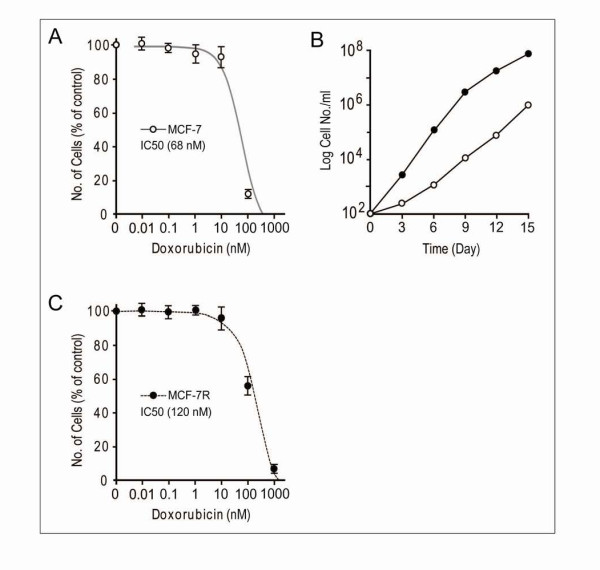
Background: Cancer cells recurrently develop into acquired resistance to the administered drugs. The iatrogenic mechanisms of induced chemotherapy-resistance remain elusive and the degree of drug resistance did not exclusively correlate with reductions of drug accumulation, suggesting that drug resistance may involve additional mechanisms. Our aim is to define the potential targets, that makes drug-sensitive MCF-7 breast cancer cells turn to drug-resistant, for the anti-cancer drug development against drug resistant breast cancer cells.

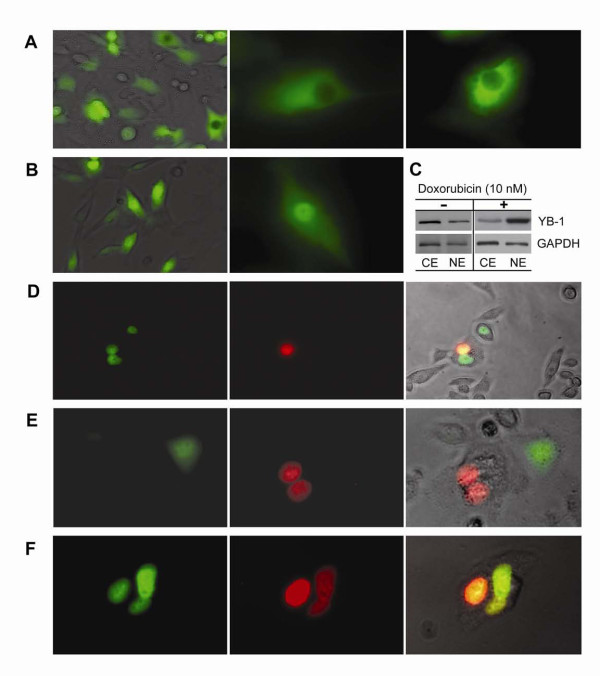
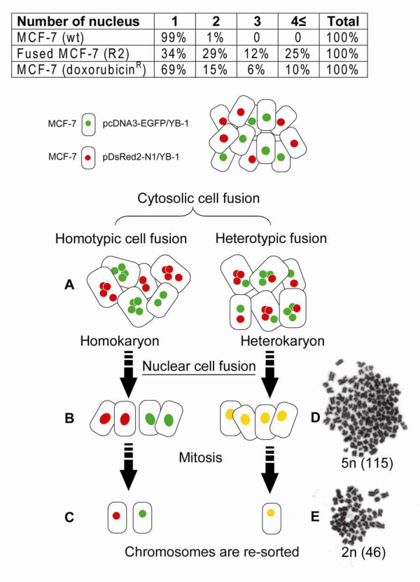
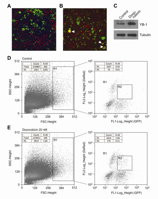
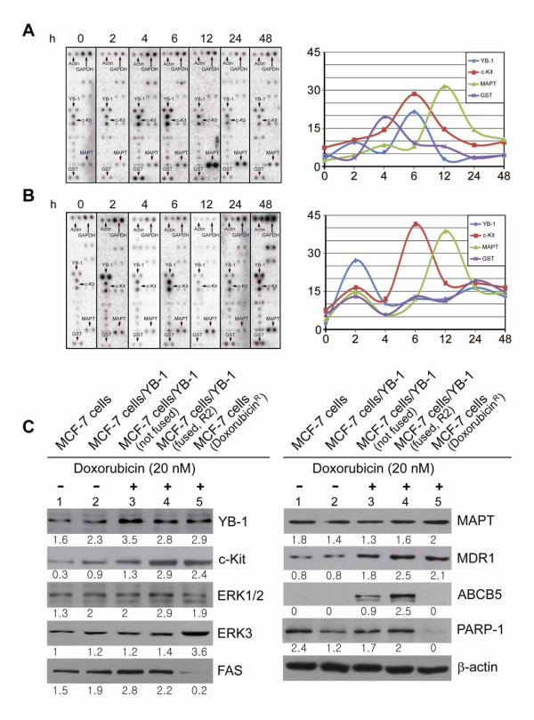
Methods: Doxorubicin resistant human breast MCF-7 clones were generated. The doxorubicin-induced cell fusion events were examined. Heterokaryons were identified and sorted by FACS. In the development of doxorubicin resistance, cell-fusion associated genes, from the previous results of microarray, were verified using dot blot array and quantitative RT-PCR. The doxorubicin-induced expression patterns of pro-survival and pro-apoptotic genes were validated.

Results: YB-1 and ABCB5 were up regulated in the doxorubicin treated MCF-7 cells that resulted in certain degree of genomic instability that accompanied by the drug resistance phenotype. Cell fusion increased diversity within the cell population and doxorubicin resistant MCF-7 cells emerged probably through clonal selection. Most of the drug resistant hybrid cells were anchorage independent. But some of the anchorage dependent MCF-7 cells exhibited several unique morphological appearances suggesting minor population of the fused cells maybe de-differentiated and have progenitor cell like characteristics.

Conclusion: Our work provides valuable insight into the drug induced cell fusion event and outcome, and suggests YB-1, GST, ABCB5 and ERK3 could be potential targets for the anti-cancer drug development against drug resistant breast cancer cells. Especially, the ERK-3 serine/threonine kinase is specifically up-regulated in the resistant cells and known to be susceptible to synthetic antagonists.


Figures

Figure 1
Figure 1
Effect of different concentration of doxorubicin treatments on the survival of MCF-7 breast cancer cell lines. A and C: Viability of MCF-7 cells in the presence of a series of concentrations of doxorubicin (0.01-1000 nM) for two weeks, the percent viability was measured following the standard protocols (non-radioactive cell proliferation assay kit, Promega). B: Control MCF-7 cells; black circlet (●), doxorubicin-treated cells; white circlet (). Time dependence effect response of doxorubicin (10 nM) on cellular growth rate of MCF-7 cells was assayed by comparing the each value with control. For the counting of colony number, cells were stained with 1% methylene blue for 20 m and washed with water. Each data point is the average of at least three independent experiments performed.
Figure 2
Figure 2
Doxorubicin-induced cell fusion. Translocation of YB-1-GFP and YB-1-Ds-Red fusion protein in respond to doxorubicin treatment. Confocal images of the two fluorescent tagged YB-1 proteins localized to each of the nuclei of non-fused MCF-7 cells and localized in one nucleus in fused MCF-7 cells. A: Control; MCF-7/YB-1-GFP transfectant without doxorubicin treatment. B--F: Demonstrate nuclear translocation of YB-1. C: Western blots of YB-1 protein from the fractionated samples; 1 × 10⁶ cells were lysed using Pierce Nuclear and Cytoplasmic Extraction Reagent Kit. 10 μg of each cytosolic extract; CE, nuclear extract; NE proteins were analyzed by 10% SDS-PAGE and Western blotted using specific antibodies diluted 1:1000 (YB-1) or 1:10000 (GAPDH). Secondary antibody (anti-mouse) diluted 1:25,000 was used with chemiluminescent substrate for detection. Cells were grown for 6 days before visualized by phase contrast and fluorescence microscopy (Axiovert 200, Carl Zeiss). D--F: MCF-7/pcDNA3-EGFP/YB-1 and MCF-7/pDsRed2-N1/YB-1 cells were mixed (1 to 1 ratio) and co-cultured with 10 nM doxorubicin and visualized by phase contrast and fluorescence microscopy. Green; GFP, Red; dsRed, Left panels; GFP detection. Middle panels; dsRed detection. D and F: Right panels; merged (yellow or orange) images represent fused MCF-7 cells.
Figure 3
Figure 3
Schema of cell fusion and chromosomal instability. A: Cytosolic cell fusion of two subtype of cells (homotype or heterotype). B and D: Most of cells, at the stage of nuclear cell fusion, revealed no further propagation and subsequently vanished. C: Viable progeny cells propagated again in culture. Giemsa-stained chromosomes of MCF-7 cells were examined. D: Abnormal sets of chromosomes as a consequence of cell fusion and (E) the cells containing giant nuclei (polyploid) were died out and the diverse populations were analogously converged into doxorubicin resistant clones. Table insert, fusion was quantitated by counting the number of cells (%) and nuclei present in a microscope field.
Figure 4
Figure 4
Cell to cell fusion occurred after doxorubicin treatment. A: MCF-7/pcDNA3-EGFP/YB-1 and MCF-7/pDsRed2-N1/YB-1 cells were co-cultured (1 to 1 ratio) without doxorubicin, (B) co-cultured with 10 nM doxorubicin. Arrow heads indicate fusion events (merged colour of green and red). C: Doxorubicin induced YB-1 overexpression; each cell lysates from (A and B) were separated by 10% SDS-PAGE, visualized by Western blot using YB-1, tubulin specific antibodies and secondary antibody (anti-mouse) with chemiluminescent substrate. D and E: FACS isolation of fused cells from the co-cultures, gated first (R1) for their size, then the gated merged colored cells (R2) were sorted. GFP fluorescence is plotted on the x-axis (FL1-Log_Height), while red fluorescence is plotted on the y-axis (FL2-Log_Height). The cells (gated R1) represent for the relatively large size of cell population. Double positive cells are gated in R2 region represent hybrids.
Figure 5
Figure 5
Differential expression patterns of YB-1, c-Kit, MAPT and GST in time course. MCF-7 cells that incubated with 20 nM doxorubicin for the indicated periods of time revealed different kinetic patterns for YB-1 and GST expressions between sensitive and resistant MCF-7 cells. A: Dot blot array analysis on doxorubicin sensitive MCF-7 cells. B: Dot blot array analysis on the doxorubicin resistant MCF-7 cells. A and B: The scale on x-axis is not in proportion with time. In the time course study, actin was employed as a control for normalization, because GAPDH was regulated in doxorubicin resistant MCF-7 cells. C: Effect of doxorubicin on the expression of drug resistance related target proteins YB-1, c-Kit, ERK1/2, ERK3, FAS, MAPT, MDR1, ABCB5 and PARP-1 in the four subtypes of MCF-7 cells. 1, 2 are MCF-7 and MCF-7/vector-YB-1 respectively without treatment of doxorubicin. 3, MCF-7/vector-YB-1 with treatment of doxorubicin for 6 h (not fused cells); 4, MCF-7/vector-YB-1 with treatment of doxorubicin for 6 h (fused cells, FACS sorted R2); 5, doxorubicin resistant MCF-7 cell line. Numbers indicate a relative level of protein expression based on the level of intensity of β-actin after normalization.

References

    1. Jasmin C, Gil-Delgado MA, Marino JP, Ecstein E, Descorps-Declere A, Misset JL. Phase I-II constant infusion of adriamycin (doxorubicin) by ambulatory pump delivery system in heavily pretreated (including adriamycin) breast cancer patients. Ann Oncol. 1990;1:189–193. - PubMed
    1. White SC, Lorigan P, Middleton MR, Anderson H, Valle J, Summers Y, Burt PA, Arance A, Stout R, Thatcher N. Randomized phase II study of cyclophosphamide, doxorubicin, and vincristine compared with single-agent carboplatin in patients with poor prognosis small cell lung carcinoma. Cancer. 2001;92:601–608. doi: 10.1002/1097-0142(20010801)92:3<601::AID-CNCR1360>3.0.CO;2-K. - DOI - PubMed
    1. Kruh GD. Introduction to resistance to anticancer agents. Oncogene. 2003;22:7262–7264. doi: 10.1038/sj.onc.1206932. - DOI - PubMed
    1. O'Driscoll L, Clynes M. Molecular markers of multiple drug resistance in breast cancer. Chemotherapy. 2006;52:125–129. doi: 10.1159/000092540. - DOI - PubMed
    1. Clarke R, Dickson RB, Brünner N. The process of malignant progression in human breast cancer. Ann Oncol. 1990;1:401–407. - PubMed

Publication types

MeSH terms