. 2010 Oct;91(4):513-23.
doi: 10.1016/j.exer.2010.07.007. Epub 2010 Jul 27.

Mapping the entire human corneal nerve architecture

Affiliations

Mapping the entire human corneal nerve architecture

Jiucheng He et al. Exp Eye Res. 2010 Oct.

Abstract

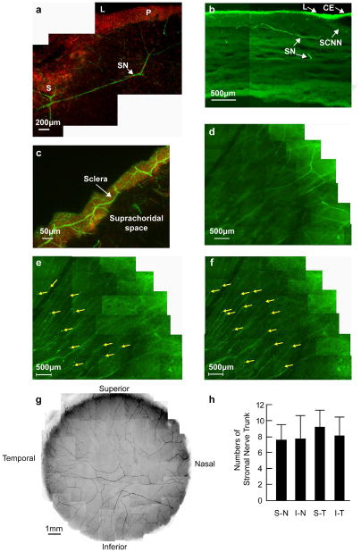
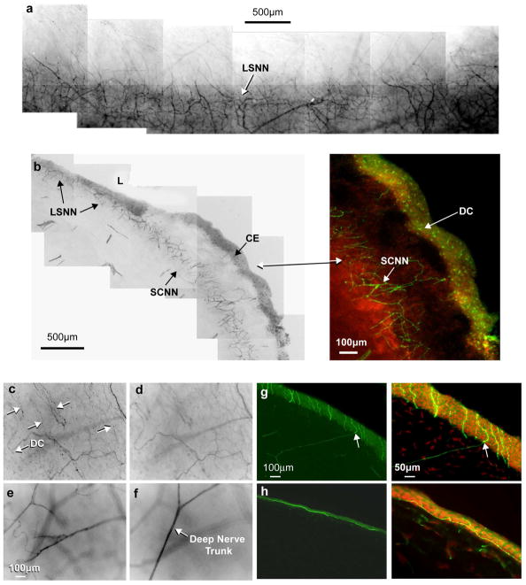
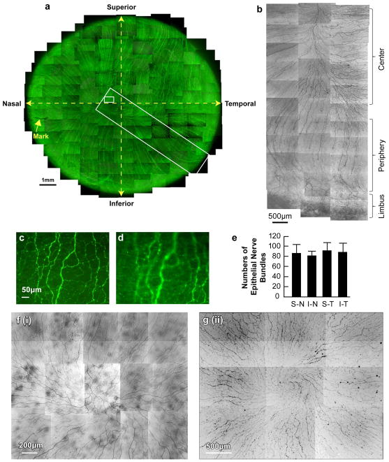
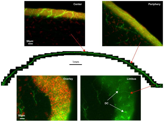
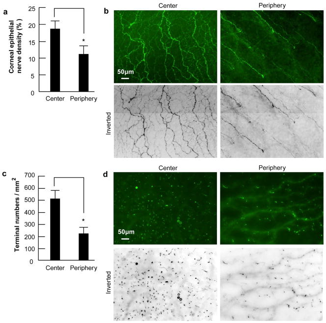
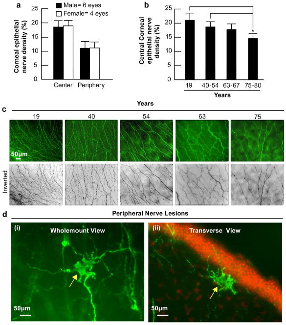
We developed an approach to generate a three-dimensional map that facilitates the assessment of epithelial nerve density in different corneal areas to define aging and gender influence on human corneal nerve architecture. Twenty-eight fresh human eyes from 14 donors of different ages were studied. Corneal nerves were stained and consecutive images acquired with a fluorescence microscope, recorded at the same plane, and merged for viewing the complete epithelial and stromal nerve architecture. After whole mount examination, the same cornea was also used for transection. Stromal nerves entered the cornea in a radial pattern, subsequently dividing into smaller branches. Some branches connected at the center of the stroma, but most penetrated upward into the epithelium. No differences were observed between nerve densities in the four corneal quadrants. Epithelial innervation in the limbal and most of the peripheral area was supplied by a superficial network surrounding the limbal area. Central epithelial nerves were supplied by branches of the stromal nerve network. Epithelial nerve density and terminal numbers were higher in the center of the cornea, rather than the periphery. There were no differences in epithelial nerve density between genders, but there was a progressive nerve density reduction concomitant with aging, mainly in eye samples of donors 70-years of age and older. The modified technique of tissue preparation used for this study allowed for observation of new nerve structure features and, for the first time, provided a complete view of the human corneal nerve architecture. Our study reveals that aging decreases the number of central epithelial nerve terminals, and increases the presence of irregular anomalies beneath the basal layer.


Figures

Figure 1
Figure 1. The origin and location of corneal stromal nerves
(a) Transverse section showing the course of nerve fibers running from the sclera to the peripheral corneal stroma. DAPI (red) was used to stain the nuclei. S: sclera; L: limbus; P: periphery; SN: stromal nerves. (b) Location of stromal nerves in the limbal area and peripheral cornea. CE: conjunctival epithelium; SCNN: Sub-conjunctival nerve network; L: limbus; SN: Stromal nerves. (c) Horizontal section showing scleral nerves along the limbus at the sclera side. (d-f) Whole mount view of corneal nerves in the limbal and peripheral cornea in the superior-temporal quadrant. (d) Deep stromal nerves, which divide into several branches upon entering the cornea. (e) Transition of stromal nerves to epithelial nerves. (f) Epithelial nerves. The tips are perforation points of the stromal nerves in the epithelium. (g) Whole mount view of human corneal stromal nerve network. The images were taken with a Nikon SMZ-1500 stereo microscope and a 4× lens from the right eye of a 45-year-old male donor. (h) Distribution of stromal nerve main branches in the four quadrants of the human corneas. The position of each quadrant was marked before removal of the cornea, as explained in Materials and methods. The number of stromal nerves from 10 corneas (ages 40–57 years old) were counted at the corneo-scleral junction with a Nikon SMZ-1500 stereo microscope. Data expressed as average ± SD.
Figure 2
Figure 2. Origin of corneal epithelial nerves
(a) Whole mount view of the limbal superficial nerve network (LSNN), which surrounds the corneal limbus and extends from the sub-conjunctival nerve network (SCNN). (b) Transected view of the LSNN and SCNN. Inset shows numerous dendritic cells (DC) present in the conjunctival epithelium (CE). (c-f) Montages of stacks showing the nerves at the limbal area at different depths. Arrows indicate the fine nerve bundles at the superficial layer and numerous DCs. (g) Epithelial nerves derived from the stromal branches. The transected view shows the perforating sites of stromal nerve branch tips (arrows). (h) Perpendicular section from the cornea of the 19–year-old donor shows the epithelial nerve bundles running horizontal in the epithelium close to the basal cells. DAPI was used to stain the nuclei.
Figure 3
Figure 3. Whole mount view of corneal epithelial nerve network
(a) Images were acquired in a time-lapse mode with a Nikon Eclipse TE200 and with a 10× lens in compliance with the natural shape of cornea obtained from the left eye of a 45-year-old male donor. More than a thousand stacks were merged together to construct the whole view of the distribution of corneal epithelial nerves. (b-d) Details of two chosen areas of the cornea. (b) Detailed course of epithelial nerve bundles running from the periphery to convergence at the center of the cornea. (c) Detailed structure of the epithelial nerve network in the center. (d) Image from the same frame as b, but with a different focus to show the epithelial nerve terminals. (e) Distribution of epithelial nerve bundles in the four quadrants. The numbers of epithelial nerve bundles were counted from seven human corneas (ages 40, 44, 45 and 54 years old) based on the whole mount view. Data expressed as average ±SD. (f). Representative patterns of convergence taken from a 54-year-old female (i) and a 63-year-old male (ii).
Figure 4
Figure 4. Transected view of the entire epithelial nerve network
For the reconstruction, the same cornea as in Figure 3 was used. Representative images from the whole view montage, counterstained with DAPI, show the features of epithelial nerves at the center and periphery. In the limbal area, numerous dendritic cells stained with βIII-tubulin are visible.
Figure 5
Figure 5. Difference of corneal epithelial nerve density and nerve terminals between the central and the peripheral zone
(a) Nerve density in human corneas was calculated as percentage of total area in each image. Eighty images for each zone from 10 corneas were used. Data expressed as average ± SD. * p< 0.001 (b) Representative images of central and peripheral epithelial nerves. (c) Number of nerve terminals. Forty images for each zone from 10 corneas were used. The terminal numbers in each image were directly counted. Because each image took up an area of 0.335 mm2, the terminal numbers per mm2 were calculated. Data expressed as average ± SD. p<0.001. (d) Representative images of nerve terminals taken from a 52–year-old male donor from the center and the periphery zones.
Figure 6
Figure 6. Effect of gender and aging in corneal epithelial nerve density
(a) Sixty-four images (32 images for each zone) from four female corneas and 96 images (48 images for each zone) from six male corneas were used. (b) Eight images were recorded from the central zone of each cornea. Forty-eight images from each group were used for analysis, except for the 19-year-old group in which there were only 16 images from the central zone. Data expressed as average ± SD. Differences of central corneal nerve densities between the groups were compared by analysis of variance (ANOVA). *p < 0.05. (c) Representative whole mount images of the central cornea from different age donors. (d) Morphology and location of peripheral neuropathies in a cornea of a 75-year-old donor. i) Whole mount view of neuropathy. ii) Transected view showing the nerve lesion localized in the anterior stroma beneath the epithelial basement membrane.

References

    1. Al-Aqaba M, Fares U, Suleman H, Lowe J, Dua HS. Architecture and distribution of human corneal nerves. Br J Ophthalmol. 2009 In press. - PubMed
    1. Attias G. Die Nerven der Hornhaut des Menschen. Albrecht von Graefes Arch Klin Exp Ophthalmol. 1912;83:207–316.
    1. Auran JD, Koester CJ, Kleiman NJ, Rapaport R, Bomann JS, Wirotsko BM, Florakis GJ, Koniarek JP. Scanning slit confocal microscopic observation of cell morphology and movement within the normal human anterior cornea. Ophthalmology. 1995;102:33–41. - PubMed
    1. Battat L, Macri A, Dursun D, Pflugfelder SC. Effects of laser in situ keratomileusis on tear production, clearance, and the ocular surface. Ophthalmology. 2001;108:1230–1235. - PubMed
    1. Belmonte C, Acosta MC, Gallar J. Neural basis of sensation in intact and injured corneas. Exp Eye Res. 2004;78:513–525. - PubMed

Publication types