. 2010 Jul 27;56(5):382-91.
doi: 10.1016/j.jacc.2010.04.019.

Impact of mechanical unloading on microvasculature and associated central remodeling features of the failing human heart

Affiliations

Impact of mechanical unloading on microvasculature and associated central remodeling features of the failing human heart

Stavros G Drakos et al. J Am Coll Cardiol. .

Abstract

Objectives: This study investigates alterations in myocardial microvasculature, fibrosis, and hypertrophy before and after mechanical unloading of the failing human heart.

Background: Recent studies demonstrated the pathophysiologic importance and significant mechanistic links among microvasculature, fibrosis, and hypertrophy during the cardiac remodeling process. The effect of left ventricular assist device (LVAD) unloading on cardiac endothelium and microvasculature is unknown, and its influence on fibrosis and hypertrophy regression to the point of atrophy is controversial.

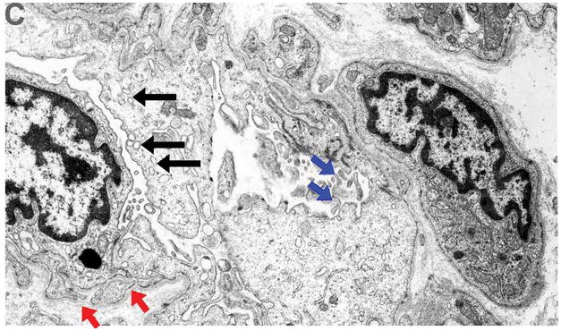
Methods: Hemodynamic data and left ventricular tissue were collected from patients with chronic heart failure at LVAD implant and explant (n = 15) and from normal donors (n = 8). New advances in digital microscopy provided a unique opportunity for comprehensive whole-field, endocardium-to-epicardium evaluation for microvascular density, fibrosis, cardiomyocyte size, and glycogen content. Ultrastructural assessment was done with electron microscopy.

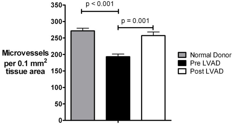
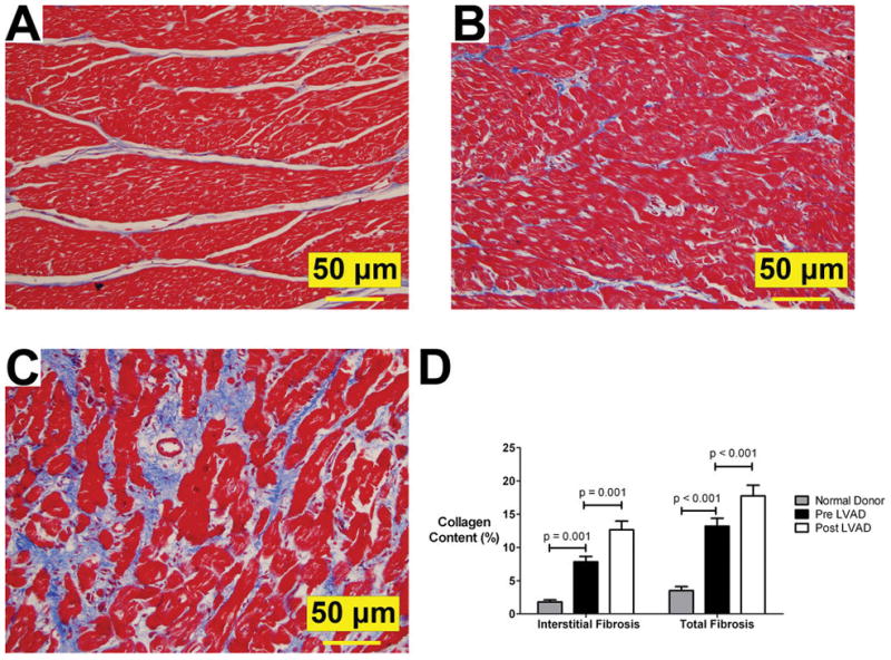
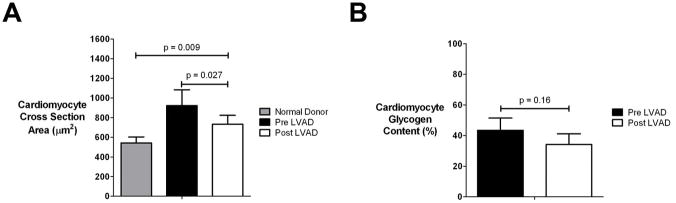
Results: Hemodynamic data revealed significant pressure unloading with LVAD. This was accompanied by a 33% increase in microvascular density (p = 0.001) and a 36% decrease in microvascular lumen area (p = 0.028). We also identified, in agreement with these findings, ultrastructural and immunohistochemical evidence of endothelial cell activation. In addition, LVAD unloading significantly increased interstitial and total collagen content without any associated structural, ultrastructural, or metabolic cardiomyocyte changes suggestive of hypertrophy regression to the point of atrophy and degeneration.

Conclusions: The LVAD unloading resulted in increased microvascular density accompanied by increased fibrosis and no evidence of cardiomyocyte atrophy. These new insights into the effects of LVAD unloading on microvasculature and associated key remodeling features might guide future studies of unloading-induced reverse remodeling of the failing human heart.


Conflict of interest statement

No conflict of interest exist

Figures

Figure 1
Figure 1. Microvasculature evaluation
Panels A and B: Left ventricular mid- myocardium immunohistochemically stained for the endothelial cell protein CD-34 (brown color). Algorithm thresholds were appropriately adjusted to allow microvascular evaluation within selected regions of interest. Panel A: 20x magnification, Panel B: 60X magnification Panel C. Automatic completion of stained microvessels by the analysis algorithm enables measurements like vessel perimeter, lumen area etc.
Figure 2
Figure 2. Collagen content evaluation
Sections were stained with Masson’s Trichrome stain- collagen stains blue. Panel A: Representative stained image from mid-myocardium before digital analysis was performed. Panel B: The section was digitally analyzed for collagen content based on color thresholds. The analysis algorithm “highlighted” collagen as dark blue and was sufficiently sensitive and accurate to exclude even small nuclei (arrows) within the fibrous tissue. Panel C: “Interstitial fibrosis” was defined as the collagen content determined in manually selected regions of interest that excluded bands of perivascular fibrosis associated with any vessel with diameter > 60μm (i.e. medium/large vessels). Panel D: Whole slide, epicardium-to-endocardium image showing superimposed all the regions selected for assessment of “interstitial fibrosis” based on the criterion described in panel C (0.5x magnification). Panel E: “Total” collagen content (“total fibrosis”) was determined by including in our analysis the whole-field stained tissue without excluding any areas (0.5x magnification).
Figure 3
Figure 3. Microvascular density
(Plots represent means ± standard error), LVAD: left ventricular assist device
Figure 4
Figure 4. Endothelial cell activation
Ultrastructural appearance (10,000x magnification, patient #2) of capillaries pre and post left ventricular assist device (LVAD) unloading revealed strong evidence of endothelial cell activation post LVAD. Panel A (pre LVAD). Red arrowheads: basal lamina, small blue arrows: cytoplasmic organelles and nuclei, big red arrow: capillary lumen. Panel B (post LVAD). Red arrowheads: basal lamina reduplication, small blue arrows: increased nuclei size and increased cytoplasm organelles protruding into the capillary lumen- irregular luminal surface (big red arrow) Panel C (post LVAD). Basal lamina reduplication (red arrows), increased nuclei and cytoplasmic size with increased pinocytotic vesicles protruding into the capillary lumen (black arrows), and numerous irregular lumenal and surface membrane projections (blue arrows), all indicative of endothelial activation.
Figure 5
Figure 5. Microvascular lumenal area
(Plots represent means ± standard error), LVAD: left ventricular assist device
Figure 6
Figure 6. Cardiac fibrosis
Panel A (normal donor heart), panel B (pre LVAD), panel C (post LVAD): Increased fibrosis (Masson’s stain- collagen content stains blue) post 63 days of LVAD-induced unloading (patient # 2); 20x magnification; Panel D: Interstitial fibrosis and Total fibrosis – see text for definitions (Plots represent means ± standard error). LVAD: left ventricular assist device
Figure 7
Figure 7. Cardiomyocyte studies
Panel A. Cardiomyocyte size evaluation Panel B. Cardiomyocyte glycogen stores evaluation (Plots represent means ± standard error), LVAD: left ventricular assist device

References

    1. Fang JC. Rise of the Machines -- Left Ventricular Assist Devices as Permanent Therapy for Advanced Heart Failure. N Engl J Med. 2009 Nov 17; [Epub ahead of print] - PubMed
    1. Klotz S, Danser AHJ, Burkhoff D. Impact of left ventricular assist device (LVAD) support on the cardiac reverse remodeling process. Prog Biophys Mol Biol. 2008;97:479–96. - PubMed
    1. Drakos SG, Terrovitis JV, Anastasiou-Nana MI, Nanas JN. Reverse remodeling during long-term mechanical unloading of the left ventricle. J Mol Cell Cardiol. 2007;43:231–42. - PubMed
    1. Katz AM. Maladaptive growth in the failing heart: the cardiomyopathy of overload. Cardiovasc Drugs Ther. 2002;16:245–9. - PubMed
    1. Soppa GK, Barton PJ, Terracciano CM, Yacoub MH. Left ventricular assist device-induced molecular changes in the failing myocardium. Curr Opin Cardiol. 2008;23:206–18. - PubMed

Publication types