. 2010 Sep 24;285(39):29911-24.
doi: 10.1074/jbc.M109.099697. Epub 2010 Jul 22.

Regulation of sealing ring formation by L-plastin and cortactin in osteoclasts

Affiliations

Regulation of sealing ring formation by L-plastin and cortactin in osteoclasts

Tao Ma et al. J Biol Chem. .

Erratum in

  • J Biol Chem. 2011 Apr 8;286(14):12812. Madayiputhiya, Nandakumar [added]

Abstract

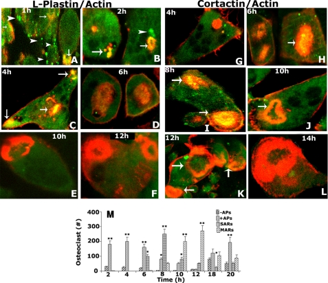
The aim of this study is to identify the exact mechanism(s) by which cytoskeletal structures are modulated during bone resorption. In this study, we have shown the possible role of different actin-binding and signaling proteins in the regulation of sealing ring formation. Our analyses have demonstrated a significant increase in cortactin and a corresponding decrease in L-plastin protein levels in osteoclasts subjected to bone resorption for 18 h in the presence of RANKL, M-CSF, and native bone particles. Time-dependent changes in the localization of L-plastin (in actin aggregates) and cortactin (in the sealing ring) suggest that these proteins may be involved in the initial and maturation phases of sealing ring formation, respectively. siRNA to cortactin inhibits this maturation process but not the formation of actin aggregates. Osteoclasts treated as above but with TNF-α demonstrated very similar effects as observed with RANKL. Osteoclasts treated with a neutralizing antibody to TNF-α displayed podosome-like structures in the entire subsurface and at the periphery of osteoclast. It is possible that TNF-α and RANKL-mediated signaling may play a role in the early phase of sealing ring configuration (i.e. either in the disassembly of podosomes or formation of actin aggregates). Furthermore, osteoclasts treated with alendronate or αv reduced the formation of the sealing ring but not actin aggregates. The present study demonstrates a novel mechanistic link between L-plastin and cortactin in sealing ring formation. These results suggest that actin aggregates formed by L-plastin independent of integrin signaling function as a core in assembling signaling molecules (integrin αvβ3, Src, cortactin, etc.) involved in the maturation process.


Figures

FIGURE 1.
FIGURE 1.
Analysis of actin distribution in osteoclasts subjected to bone resorption. Osteoclasts plated on dentine slices for 3 h (A and A′), 8 h (B), and 12 h (C) were stained with rhodamine-phalloidin to determine the changes in the distribution of actin during bone resorption. Osteoclasts are outlined in A′ to indicate stretching off of membrane at 2–3 h. Actin aggregates that most likely function as secondary adhesive structures are indicated by arrows in A′. An increase in the number of actin rings (B and C) was observed as a result of the increased surface area of osteoclasts at 8 and 12 h. Scale bars, 25 μm. The results are representative of four experiments with four separate osteoclast preparations.
FIGURE 2.
FIGURE 2.
Analysis of cell shape changes in osteoclasts incubated with native long bone particles. A, bone particles (∼60–80 μm in size) isolated from the long bones of mice. B, confluent osteoclast in culture prior to bone treatment is shown. Osteoclasts were incubated with bone particles (60–80 μm size) for 4 h (C) and 6 h (D). The arrowheads in C and D indicate osteoclasts that are attached to bone particles. These osteoclasts are capable of forming actin ring and bone resorption. Osteoclasts were viewed under a ×40 objective in an inverted microscope. Experiments were repeated with more than 10 different osteoclast preparations. The data shown are representative of those experiments.
FIGURE 3.
FIGURE 3.
Analysis of the expression profile of proteins in osteoclasts treated with and without native bone particles. A, treatments of osteoclasts with bone particles were performed as described under “Experimental Procedures.” Lanes 1 and 2, lysate proteins made from osteoclasts incubated with (+) and without (−) bone particles for 16–18 h were subjected to 10% SDS-PAGE and Coomassie Blue staining analyses. Lanes 3 and 4, immunoblotting analysis with a cortactin (top) and L-plastin antibody (bottom). B, osteoclasts were treated with bone particles for different time periods as indicated at the bottom. Lysates were separated on SDS-polyacrylamide gels and then transferred and probed with L-plastin and actin antibodies sequentially without stripping. C, equal amounts of lysate proteins from osteoclasts treated as indicated in the figure were subjected to SDS-PAGE and Coomassie Blue staining. (−), untreated osteoclasts; BP, bone particles; OD, osteologic discs; DP, dentine particles. D, equal amounts of protein lysates made from osteoclasts treated with (lanes 2 and 4) and without (lanes 1 and 3) bone particles were subjected to three SDS-10% polyacrylamide gels. Blots made from these three gels were immunoblotted with anti-Tyr(P) (p-Tyrosine), p130cas, and PYK2 antibody, respectively. These blots were stripped and blotted correspondingly with GAPDH/cortactin, WASP, and Src antibody. Immunoblotting of the first blot with GAPDH (37 kDa) and cortactin (80–85 kDa) antibody was performed sequentially without any further stripping. E, immunoblotting (IB) analysis with a Ser(P) (p-serine) antibody. This immunoblot was sequentially probed with a Ser(P), L-plastin, and GAPDH antibody. Immunoblotting analysis with a GAPDH (A, D, or E) or actin (B) antibody was used as a loading control. The results are representative of three experiments performed with three separate osteoclast preparations.
FIGURE 4.
FIGURE 4.
SDS-PAGE and immunoblotting analyses of lysates made from osteoclasts subjected to various treatments. A and B, time-dependent effects of TNF-α on L-plastin and cortactin levels. Equal amounts of lysate proteins were used for immunoblotting analysis with an antibody (ab) to L-plastin (A) and cortactin (B). Actin was used as a loading control in A and B. C, equal amounts of lysate proteins from osteoclasts treated as indicated in the figure were subjected to SDS-PAGE (10%) and Coomassie Blue staining analyses. MCSF, macrophage colony-stimulating factor; (−) and (+), untreated and bone particle-treated; IgG, control nonspecific IgG control; TNFR1, anti-TNF-1 receptor. D and E, equal amount of lysate proteins from osteoclasts treated with various treatments, as indicated in the figure, were used for SDS-PAGE/Coomassie Blue staining (D) and immunoblotting analyses (E). Immunoblotting analyses with phosphocortactin (Tyr(P)421) (E, top, p-Cortactin), cortactin (E, middle), and GAPDH (E, bottom) are shown. F–I, bone resorption assay in vitro. Osteoclasts were treated as indicated at the top of each panel (E–F). Pits were scanned under confocal microscopy. Resorption pits were seen as dark spots. Scale bar, 25 μm. These results represent one of three separate experiments performed with the same results.
FIGURE 5.
FIGURE 5.
Confocal analysis of time-dependent changes in actin organization and localization of L-plastin and cortactin in resorbing osteoclasts. A–L, confocal microscopic images of osteoclasts stained for actin (red), L-plastin (green; A–F), and cortactin (green; G–L) are shown. Yellow color (indicated by arrows) represents the colocalization of proteins (A–C and H–J). M, changes in actin organization were determined in ∼300 osteoclasts and provided as a graph. Measurements were performed at the indicated time below the graph. Shown is basic actin staining with no actin patches or aggregates (−APs), with actin patches or aggregates (+APs), with small actin rings (SARs), and with multiple or single big mature actin rings (MARs). Data are provided as mean ± S.E. of three experiments. **, p < 0.001; *, p < 0.05 versus cells with no actin patches.
FIGURE 6.
FIGURE 6.
Analysis of the effects of depletion or knockdown of cortactin on sealing ring formation and bone resorption. I, immunoblotting analysis of cortactin levels in osteoclasts treated with control RNAi (Sc; 200 nm) and siRNA (Si; 100 and 200 nm) to cortactin (A). Immunoblotting analysis with GAPDH is shown in B. II, the effects of ScRNAi (A and D) and siRNA (B, C, and E) to cortactin on sealing ring formation. Osteoclasts were stained for cortactin (green) and actin (red). Cortactin distribution is shown in A–C. The effects of scrambled RNAi and SiRNA on multiple osteoclasts are shown in D and E. Osteoclasts were stained for actin (red) with rhodamine-phalloidin. Dentine is shown by the reflected light (green in D and E). Resorption pits were found underneath osteoclasts (D, D″, E, and E″). Actin distribution (D′ and E′) and resorption lacuna (D″ and E″) are shown separately in gray. Actin aggregates and superficial pits are indicated by asterisks in E and E″. Sealing rings are indicated by arrowheads (D and D′) and arrows (E and E′). Scale bar, 25 μm. III, bone resorption assay in vitro. Osteoclasts treated with scrambled ScRNAi and siRNA were cultured on dentine for 24 h. Pits were scanned in a confocal microscopy. Resorption pits were seen as dark spots. Experiments were repeated three times with three different osteoclast preparations. These results represent one of three separate experiments performed with the same results.
FIGURE 7.
FIGURE 7.
I, confocal analysis of localization of actin with cortactin and integrin β3. Osteoclasts cultured on dentine slices for 12–14 h were processed for double staining with actin/cortactin (I, A and B) and actin/integrin β3 (C). Confocal microscopic images of osteoclasts stained for actin (A–C, red), cortactin (A′, green), and integrin β3 (C′, green) are shown. Yellow color represents colocalization of cortactin and integrin β3 with actin (overlay panels A″ and C″). A higher magnification view of the area indicated by an arrow in A″ is shown in B and B″. More than eight actin rings (outlined in B″) were observed in this osteoclast. Resorption pits are indicated by asterisks, and an osteoclast that exhibits single or multiple sealing rings is indicated by an arrowhead (A, C, and C″). Scale bars, 10 μm (B) and 25 μm (C″). II, confocal analysis of localization of actin with cortactin, integrin β3, and L-plastin in osteoclasts treated with siRNA and ScRNAi to cortactin. Osteoclasts were treated with ScRNAi (A) and siRNA to cortactin (B and C). Confocal microscopic images of osteoclasts stained for actin (A–C, red), cortactin (A, green), integrin β3 (B, green), and L-plastin (C, green) are shown. Yellow color represents colocalization of cortactin and integrin β3 with actin (overlay panels A and B). Scale bar, 25 μm. The results represent one of the three separate experiments performed with three different osteoclast preparations.
FIGURE 8.
FIGURE 8.
I, confocal analysis of time-dependent changes in actin organization and bone resorption in response to TNF-α. Osteoclasts plated on dentine slices were treated with TNF-α for 2–3 h (A), 8 h (B), and 12 h (C). Confocal microscopic images of osteoclasts stained for actin are shown. Distribution of actin (red) and scans of dentine slices (green) are shown separately. Several resorption pits were observed after incubation with TNF-α for 12–14 h. II, confocal analysis of the effects of various treatments on sealing ring formation and bone resorption. Osteoclasts were treated with a neutralizing antibody to TNF-α (B), Src (C), and αv inhibitor (D) in the presence of TNF-α. Osteoclasts treated together with a species-specific IgG and TNF-α were used as control (A) for neutralizing antibody to TNF-α antibody treatment (B). Sealing rings are indicated by arrowheads in A. Punctate podosome-like structures are indicated by crooked arrows in B and C (red panel). Actin patches (B and C, red panel) and aggregates are indicated by arrows (D, red panel). Bone resorption is considerably reduced in osteoclasts treated with a neutralizing antibody to TNF-α (B) and inhibitors to Src and αv (C and D) in the presence of TNF-α (green panels). Scale bar, 25 μm. The results represent one of the three separate experiments performed with three different osteoclast preparations.

Similar articles

Cited by

References

    1. Chellaiah M., Kizer N., Silva M., Alvarez U., Kwiatkowski D., Hruska K. A. (2000) J. Cell Biol. 148, 665–678 - PMC - PubMed
    1. Babb S. G., Matsudaira P., Sato M., Correia I., Lim S. S. (1997) Cell Motil. Cytoskeleton 37, 308–325 - PubMed
    1. Hurst I. R., Zuo J., Jiang J., Holliday L. S. (2004) J. Bone Miner. Res. 19, 499–506 - PubMed
    1. McMichael B. K., Kotadiya P., Singh T., Holliday L. S., Lee B. S. (2006) Bone 39, 694–705 - PubMed
    1. Chellaiah M. A. (2005) J. Biol. Chem. 280, 32930–32943 - PubMed

Publication types

MeSH terms