. 2010 Nov;24(11):4420-32.
doi: 10.1096/fj.10-158477. Epub 2010 Jul 22.

The carboxy-terminal fragment of inhibitor-2 of protein phosphatase-2A induces Alzheimer disease pathology and cognitive impairment

Affiliations

The carboxy-terminal fragment of inhibitor-2 of protein phosphatase-2A induces Alzheimer disease pathology and cognitive impairment

Xiaochuan Wang et al. FASEB J. 2010 Nov.

Abstract

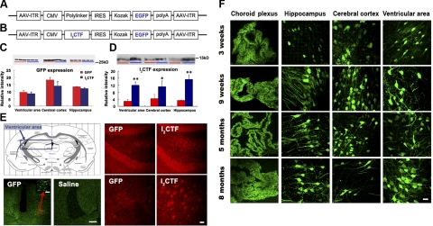
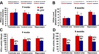
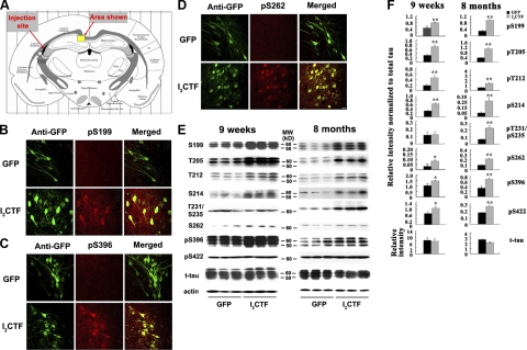
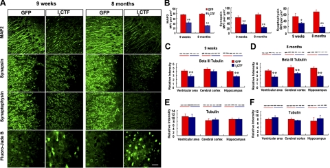
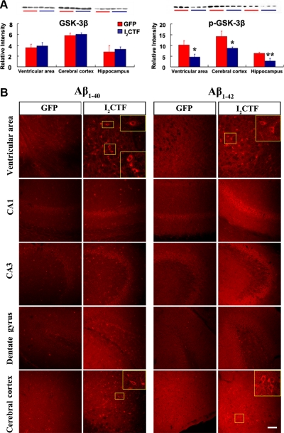
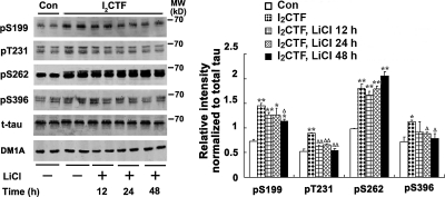
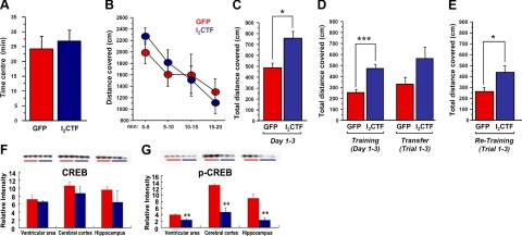
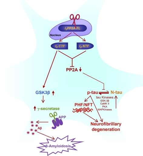
Development of rational therapeutic treatments of Alzheimer disease (AD) requires the elucidation of the etiopathogenic mechanisms of neurofibrillary degeneration and β-amyloidosis, the two hallmarks of this disease. Here we show, employing an adeno-associated virus serotype 1 (AAV1)-induced expression of the C-terminal fragment (I(2CTF)) of I(2)(PP2A), also called SET, in rat brain, decrease in protein phosphatase 2A (PP2A) activity, abnormal hyperphosphorylation of tau, and neurodegeneration; littermates treated identically but with vector only, i.e., AAV1-enhanced green fluorescent protein (GFP), served as a control. Furthermore, there was an increase in the level of activated glycogen synthase kinase-3β and enhanced expression of intraneuronal Aβ in AAV1-I(2CTF) animals. Morris water maze behavioral test revealed that infection with AAV1-I(2CTF) induced spatial reference memory and memory consolidation deficits and a decrease in the brain level of pSer133-CREB. These findings suggest a novel etiopathogenic mechanism of AD, which is initiated by the cleavage of I(2)(PP2A), producing I(2CTF), and describe a novel disease-relevant nontransgenic animal model of AD.


Figures

Figure 1.
Figure 1.
AAV1-induced gene expression in brain is stable and long lasting when newborn rats are infected intracerebroventricularly. A, B) Linear maps of the AAV vector plasmids (based on pTRUF12) used in the present study. With the exception of the inverted terminal repeats (ITR), all viral genes had been removed and replaced with GFP (A), or I2CTF and GFP (B). CMVe-CBA (cytomegalovirus enhancer-chicken beta actin promoter); IRES (internal ribosomal entry site) from poliovirus. C) Western blots developed with mAb to GFP showed a similar level of expression of GFP between AAV1-I2CTF and AAV1-GFP rats. D) In contrast, blots developed with pAb to I2CTF showed a marked increase in the brain level of I2CTF in AAV1-I2CTF as compared with AAV1-GFP animals. E) AAV1-I2CTF animals (13 wk) showed an increase in the expression of I2CTF over endogenous I2PP2A seen in AAV1-GFP rats; cytoplasmic staining of neurons of the dentate gyrus hilus (anti-I2CTF, red); fluorescence of GFP (anti-GFP, green); ventricular area studied (framed). Animals injected with saline as expression control showed only background staining. F) Immunohistofluorescence using mAb GFP showed that the AAV1-induced expression was long lasting in the neuronal cell layers of choroid plexus, hippocampus, cerebral cortex, and ventricular area. Scale bars = 400 μm (E); 80 μm (E, inset); 25 μm (F).
Figure 2.
Figure 2.
Infection with AAV1-I2CTF results in the decrease of PP2A activity without any significant change in its level. Coimmunoprecipitation was carried out on lysates of cerebral cortex, ventricular area, and hippocampus from I2CTF and, as control, GFP rats at 9 wk (A, C) and 8 mo (B, D) after infection with AAV. Lysates were immunoprecipitated with anti-PP2Ac rabbit polyclonal antibody R123d. Immunoprecipitates were divided into two parts: one part was employed for Western blots probed with anti-PP2Ac mouse monoclonal 1D6 (A, B), while the other part was used for assaying PP2Ac activity (C, D); the PP2A activity was decreased in AAV1-I2CTF in comparison to AAV1-GFP animals. *P < 0.05; **P < 0.01.
Figure 3.
Figure 3.
I2CTF induces abnormal hyperphosphorylation of tau. AD) At 9 wk postinfection, an immunohistochemical staining in a selected area of interest (A) shows expression in GFP and I2CTF rats; pS199 (B), pS396 (C), and pS262 (D) show only background in AAV1-GFP rat brain and abnormal hyperphosphorylation of tau in AAV1-I2CTF rat brain and a colocalization with I2CTF in I2CTF- but not in GFP-rats. Scale bars = 25 μm. E) Western blots show increase in abnormal hyperphosphorylation of tau in 9-wk and 8-mo I2CTF rats. F) Quantitative analysis of the blots showing levels of phosphorylated taus normalized with total tau levels (left panel) shows significant increase in tau hyperphosphorylation at several sites studied, and this increase was further enhanced from 9 wk to 8 mo (right panel) at several sites. *P < 0.05; **P < 0.01.
Figure 4.
Figure 4.
I2CTF induces neurodegeneration and loss of dendritic and synaptic plasticity. A, B) As compared with GFP rats, I2CTF rats show a marked decrease in the expression of somato-dendritic marker MAP2 and synaptic markers synapsin and synaptophysin in the hippocampus, and this decrease was enhanced from 9 wk to 8 mo post-AAV1-I2CTF infection (B). Fluoro Jade staining shows a selective neurodegeneration in the AAV1-I2CTF rat hippocampus (A). CF) Levels of the neuron-specific βΙΙΙ tubulin (C, D) and not total tubulin (E, F) were selectively decreased in ventricular area of 9-wk and all areas of the 8-mo AAV1-I2CTF rats studied. *P < 0.05; **P < 0.01.
Figure 5.
Figure 5.
I2CTF induces a selective increase in GSK-3β activity by decreasing the level of the inactive form pSer GSK-3β and an increase in the intraneuronal expression of Aβ. A) Western blots and levels of total GSK-3β and the inactive form pSer9 GSK-3β determined from the blots. Decrease in the pSer GSK-3β but not in total GSK-3β in I2CTF rats suggest an increase in the activity of this kinase. B) Immunohistochemical staining with rabbit polyclonal antibodies to Aβ34–40 and to Aβ36–42 showing a selective increase in the intraneuronal expression of Aβ. Scale bar = 100 μm.
Figure 6.
Figure 6.
I2CTF-induced tau hyperphosphorylation involves both PP2A and GSK-3β. HEK293/tau cells were transfected with pcDNA3.1-I2CTF or, as a control, with pcDNA3.1. After 6 h of transfection, the cells were treated with 20 mM lithium chloride to inhibit GSK-3 for 12 h, 24 h, and 48 h. Western blots of lysates of these cells were analyzed for phosphorylation of tau at Ser-199, Thr231, Ser-262, and Ser-396. A partial inhibition of tau phosphorylation at Ser-199, Thr231, and Ser-396, the GSK-3β-acting sites and no inhibition at Ser-262, a PP2A, but not GSK-3β site, demonstrates involvement of both PP2A and GSK-3β activities in I2CTF-induced abnormal hyperphosphorylation of tau. *P < 0.05, **P < 0.01 vs. control; ΔP < 0.05, ΔΔP < 0.01 vs. I2CTF.
Figure 7.
Figure 7.
Expression of I2CTF impairs spatial learning and memory, and this impairment is associated with a decrease in the brain level of pSer133-CREB. AD) While, as compared with the GFP rats (control group), the I2CTF rats showed no change in anxiety (A) or exploration (B) in the open field, these animals were impaired in spatial learning and memory at age 5 mo; P < 0.005 (C) and 8 mo; P < 0.01 (D, left bars) in Morris water maze during training but not in the transfer test (D, right bars). E) I2CTF animals also showed impairment in the consolidation of memory on retesting 1 mo after the previous water maze test; P < 0.05. F, G) These impairments in I2CTF rats were associated with a selective decrease in the brain level of pSer133-CREB.
Figure 8.
Figure 8.
A proposed novel etiopathogenic mechanism of AD, which is initiated by the cleavage of I2PP2A/SET into I2NTF and I2CTF.

Similar articles

Cited by

References

    1. Iqbal K., Flory M., Khatoon S., Soininen H., Pirttila T., Lehtovirta M., Alafuzoff I., Blennow K., Andreasen N., Vanmechelen E., Grundke-Iqbal I. (2005) Subgroups of Alzheimer's disease based on cerebrospinal fluid molecular markers. Ann. Neurol. 58, 748– 757 - PubMed
    1. Alafuzoff I., Iqbal K., Friden H., Adolfsson R., Winblad B. (1987) Histopathological criteria for progressive dementia disorders: clinical-pathological correlation and classification by multivariate data analysis. Acta Neuropathol. (Berl.). 74, 209– 225 - PubMed
    1. Grundke-Iqbal I., Iqbal K., Tung Y. C., Quinlan M., Wisniewski H. M., Binder L. I. (1986) Abnormal phosphorylation of the microtubule-associated protein tau (tau) in Alzheimer cytoskeletal pathology. Proc. Natl. Acad. Sci. U. S. A. 83, 4913– 4917 - PMC - PubMed
    1. Grundke-Iqbal I., Iqbal K., Quinlan M., Tung Y. C., Zaidi M. S., Wisniewski H. M. (1986) Microtubule-associated protein tau. A component of Alzheimer paired helical filaments. J. Biol. Chem. 261, 6084– 6089 - PubMed
    1. Bennecib M., Gong C. X., Grundke-Iqbal I., Iqbal K. (2000) Role of protein phosphatase-2A and -1 in the regulation of GSK-3, cdk5 and cdc2 and the phosphorylation of tau in rat forebrain. FEBS Lett. 485, 87– 93 - PubMed

Publication types

MeSH terms