. 2010 Sep;177(3):1187-97.
doi: 10.2353/ajpath.2010.091289. Epub 2010 Jul 22.

Neurovascular protective effect of FeTPPs in N-methyl-D-aspartate model: similarities to diabetes

Affiliations

Neurovascular protective effect of FeTPPs in N-methyl-D-aspartate model: similarities to diabetes

Mohammed M H Al-Gayyar et al. Am J Pathol. 2010 Sep.

Erratum in

  • Am J Pathol. 2015 Jun;185(6):1795-6

Expression of concern in

  • Note of Concern.
    [No authors listed] [No authors listed] Am J Pathol. 2016 Oct;186(10):2769. doi: 10.1016/j.ajpath.2016.07.007. Am J Pathol. 2016. PMID: 27658715 Free PMC article.

Abstract

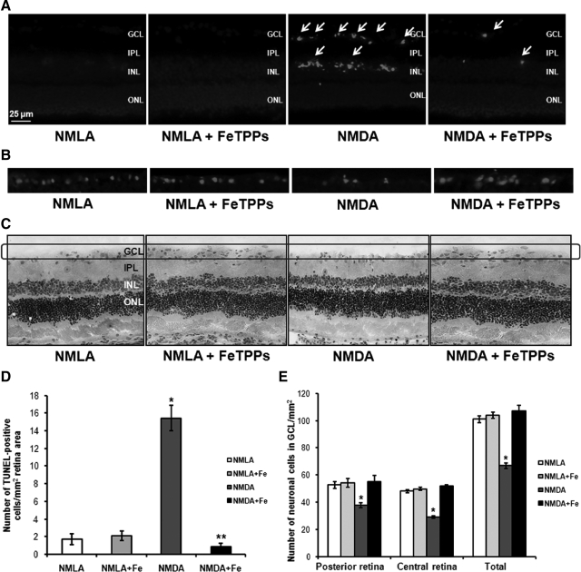
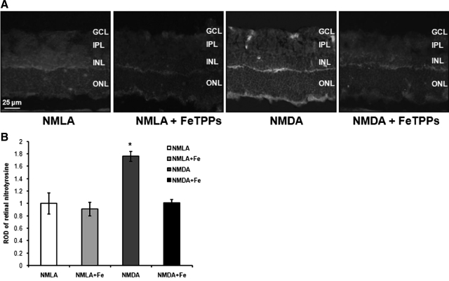
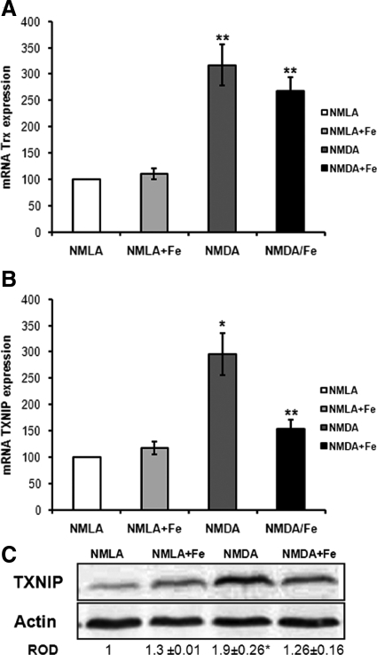
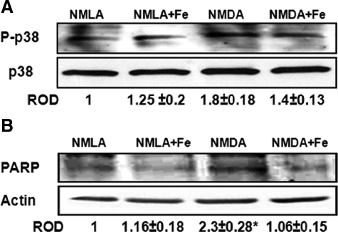
We have previously shown a causal role of peroxynitrite in mediating retinal ganglion cell (RGC) death in diabetic and neurotoxicity models. In the present study, the role of peroxynitrite in altering the antioxidant and antiapoptotic thioredoxin (Trx) system will be investigated as well as the subsequent effects on glial activation and capillary degeneration. Excitotoxicity of the retina was induced by intravitreal injection of N-methyl-d-aspartate (NMDA) in rats, which also received the peroxynitrite decomposition catalyst FeTPPs. RGC loss was assessed by terminal deoxynucleotidyl transferase-mediated dUTP nick-end labeling and GC count. Glial activation and nitrotyrosine were assessed by immunohistochemistry. Acellular capillaries and pericytes were counted in retinal trypsin digest. NMDA-induced peroxynitrite formation caused RGC loss, which was associated with enhanced expression of Trx and its endogenous inhibitor thioredoxin interacting protein. The results also showed enhanced thioredoxin interacting protein/Trx binding and disruption of the Trx/apoptosis signal-regulating kinase 1 "inhibitory complex," leading to release of apoptosis signal-regulating kinase 1 and activation of the apoptotic pathway, as evidenced by p38 MAPK and poly-ADP-ribose polymerase activation. Furthermore, NMDA caused glial activation and compromised retinal vasculature, as indicated by acellular-capillary formation and pericyte loss. Treatment with FeTPPs blocked these effects. In conclusion, NMDA-induced retinal neuro/vascular injury is mediated by peroxynitrite-altered Trx antioxidant defense, which in turn activates the apoptosis signal-regulating kinase-1 apoptotic pathway. In addition to acute RGC death, an NMDA model can be a useful tool to study glial activation and capillary degeneration in retinal neurodegenerative disorders, including diabetic retinopathy.


Figures

Figure 1
Figure 1
Decomposing peroxynitrite protected retinal ganglion cells in NMDA-injected retinas. A and D: Representative images and statistical analysis of TUNEL assay of rat retina after one day of the injection showing significant increase in number of TUNEL positive cells in NMDA-injected rats (n = 5–7, P < 0.05), especially GCL as compared with NMLA controls or NMDA + FeTPPs. The number of TUNEL positive cells (arrows) were counted in four fields of different areas of each retina and calculated as the number/mm2 of retinal area (magnification, ×200). B: Representative images of rat retina sections stained with antiBrn-3a, specific RGC marker, showing reduced number of RGC in NMDA-injected retinas as compared with NMLA controls. C and E: Representative images and statistical analysis of rat retina sections stained with H&E showing a reduction in number of GCL in NMDA-injected rats (magnification, ×200) as compared with NMLA controls or NMDA + FeTPPs in central and posterior retina (n = 4–6, P < 0.05). FeTPPs blocked all these effects in NMDA-injected retinas but did not affect NMLA control. There was a significant difference as compared with the rest of the groups at *P < 0.05. There was a significant difference as compared with the control groups at **P < 0.05. GCL, ganglion cell layer; ONL, outer nuclear layer; IPL, inner plexiform layer; INL, inner nuclear layer.
Figure 2
Figure 2
FeTPPs blocked nitrotyrosine in NMDA-injected retinas. A: Representative images showing NMDA-stimulated peroxynitrite formation in rat retina indicated by its footprint nitrotyrosine (magnification, ×200) after one day of intravitreal NMDA injection. FeTPPs blocked this effect in NMDA-injected retinas but did not affect NMLA controls. B: Statistical analysis showed 1.8-fold increase in ROD of NMDA-injected retinas as compared with NMLA controls or NMDA + FeTPPs (n = 6). There was a significant difference as compared with the rest of the groups at *P < 0.05. GCL, ganglion cell layer; ONL, outer nuclear layer; IPL, inner plexiform layer; INL, inner nuclear layer.
Figure 3
Figure 3
FeTPPs reduced NMDA-induced TXNIP but not Trx expression in NMDA-injected retinas. A: One day post NMDA injection, real-time PCR of retinal lysate showed a threefold increase in Trx expression in NMDA-injected retinas that was maintained by FeTPPs treatment as compared with NMLA controls (n = 5). B: Real-time PCR of retinal lysate showed a threefold increase in TXNIP expression in NMDA-injected retinas compared with NMLA controls (n = 5). C: Western blot of rat retinal lysates showed 1.9-fold increase in TXNIP in NMDA-injected retinas compared with NMLA control (n = 5–7). FeTPPs (100 μg/eye) blocked TXNIP expression in NMDA-injected rats but did not affect its expression in NMLA controls. There was a significant difference as compared with rest of the groups at *P < 0.05. There was a significant difference as compared with controls at **P < 0.05.
Figure 4
Figure 4
Increased TXNIP bind Trx and released ASK-1 from inhibitory complex. A: Immunoprecipitation of Trx and immunoblotting with ASK-1 showed 65% reduction in interaction between Trx and ASK-1 in NMDA-injected rats after one day as compared with NMLA controls (n = 4–5). B: Immunoprecipitation of Trx and immunoblotting with TXNIP showed a 1.7-fold increase in interaction between Trx and TXNIP in NMDA-injected retinas as compared with NMLA control (n = 4). C: Western blot analysis showed a 1.9-fold increase in ASK-1 expression in rat retina lysate of NMDA-injected retinas as compared with the NMLA controls (n = 4–6). D: Immunolocalization of ASK-1 in retinal ganglion cell and inner retinal layers in NMDA-injected retinas compared with NMLA controls (magnification, ×200). FeTPPs blocked all these effects in NMDA-injected retinas but did not affect NMLA controls. There was a significant difference as compared with the rest of the groups at *P < 0.05. GCL, ganglion cell layer; ONL, outer nuclear layer; IPL, inner plexiform layer; INL, inner nuclear layer.
Figure 5
Figure 5
FeTPPs blocked activation of p38 MAPK apoptosis pathway. A: After one day of intravitreal NMDA injection, Western blot analysis showed 1.8-fold increase in p38 MAPK phosphorylation in rat retina lysate of NMDA-injected retinas compared with NMLA controls (n = 5–6). B: Western blot analysis of rat retina lysate showed a 2.3-fold increase in cleaved PARP expression in NMDA-injected retinas compared with NMLA controls (n = 4). Treatment of animals with FeTPPs (100 μg/eye) reversed these effects in NMDA-injected retinas but did not affect NMLA controls. There was a significant difference as compared with rest of the groups at *P < 0.05.
Figure 6
Figure 6
FeTPPs prevented NMDA-induced glial activation, capillary degeneration and pericytes loss. A: Representative images of GFAP immunofluorescence in various treatment groups after one day. In NMDA-injected retinas, the end feet of the Müller cells showed abundant GFAP immunofluorescence (red), and the radial processed stained intensely throughout both the inner and the outer retina (magnification, ×400). Similar morphological changes were observed in 3–4 animals. B: Representative image of trypsin-digest stained with periodic acid-Schiff and hematoxylin showing acellular capillaries (arrows) in NMDA-injected rats after seven days (magnification, ×400). C: Representative images of acellular capillaries using confocal microscopy of trypsin digest. All endothelial cells are labeled with isolectin (red) while collagen IV (green) labels vascular basement membrane. The circle superimposed on the merged image demonstrates a typical acellular capillary in retina (collagen IV positive but isolectin negative). D: Statistical analysis showing 2.6-fold increase in number of acellular capillaries in NMDA-injected retinas compared with NMLA controls or NMDA + FeTPPs (n = 4–6). E–F: Statistical analysis and representative image of retinal trypsin-digest stained with periodic acid-Schiff (PASH) and Hematoxylin showing pericyte cells (indicated by red arrows) in NMDA-injected rats showing a significant decrease in pericytes count in NMDA-injected retinas compared with NMLA-controls or NMDA + FeTPPs (n = 4−6) (magnification, ×400). These effects were blocked by FeTPPs in NMDA-injected retinas but did not alter NMLA-controls. G: Representative image of retinal trypsin digest stained with TUNEL (left) showing that vascular cell death occurs by apoptosis as indicated by TUNEL positive nuclei (white arrows) in endothelial cells (red arrows) (400× magnification). There was significant difference against the controls and NMDA + FeTPPs groups at *P < 0.05. There was significant difference against the rest of the groups at #P < 0.05. GCL, ganglion cell layer; IPL, inner plexiform layer; INL, inner nuclear layer; ONL, outer nuclear layer.

References

    1. El-Remessy AB, Khalil IE, Matragoon S, Abou-Mohamed G, Tsai NJ, Roon P, Caldwell RB, Caldwell RW, Green K, Liou GI. Neuroprotective effect of (-)Delta9-tetrahydrocannabinol and cannabidiol in N-methyl-D-aspartate-induced retinal neurotoxicity: involvement of peroxynitrite. Am J Pathol. 2003;163:1997–2008. - PMC - PubMed
    1. Zheng L, Gong B, Hatala DA, Kern TS. Retinal ischemia and reperfusion causes capillary degeneration: similarities to diabetes. Invest Ophthalmol Vis Sci. 2007;48:361–367. - PubMed
    1. Ali TK, Matragoon S, Pillai BA, Liou GI, El-Remessy AB. Peroxynitrite mediates retinal neurodegeneration by inhibiting NGF survival signal in experimental and human diabetes. J Diabetes. 2008;57:889–898. - PubMed
    1. El-Remessy AB, Tang Y, Zhu G, Matragoon S, Khalifa Y, Liu EK, Liu JY, Hanson E, Mian S, Fatteh N, Liou GI. Neuroprotective effects of cannabidiol in endotoxin-induced uveitis: critical role of p38 MAPK activation. Mol Vis. 2008;14:2190–2203. - PMC - PubMed
    1. Schmidt KG, Bergert H, Funk RH. Neurodegenerative diseases of the retina and potential for protection and recovery. Curr Neuropharmacol. 2008;6:164–178. - PMC - PubMed

Publication types

MeSH terms

Substances