. 2010 Sep;177(3):1298-310.
doi: 10.2353/ajpath.2010.091210. Epub 2010 Jul 22.

Antibody treatment promotes compensation for human cytomegalovirus-induced pathogenesis and a hypoxia-like condition in placentas with congenital infection

Affiliations

Antibody treatment promotes compensation for human cytomegalovirus-induced pathogenesis and a hypoxia-like condition in placentas with congenital infection

Ekaterina Maidji et al. Am J Pathol. 2010 Sep.

Abstract

Human cytomegalovirus (HCMV) is the major viral cause of birth defects worldwide. Affected infants can have temporary symptoms that resolve soon after birth, such as growth restriction, and permanent disabilities, including neurological impairment. Passive immunization of pregnant women with primary HCMV infection is a promising treatment to prevent congenital disease. To understand the effects of sustained viral replication on the placenta and passive transfer of protective antibodies, we performed immunohistological analysis of placental specimens from women with untreated congenital infection, HCMV-specific hyperimmune globulin treatment, and uninfected controls. In untreated infection, viral replication proteins were found in trophoblasts and endothelial cells of chorionic villi and uterine arteries. Associated damage included extensive fibrinoid deposits, fibrosis, avascular villi, and edema, which could impair placental functions. Vascular endothelial growth factor and its receptor fms-like tyrosine kinase 1 (Flt1) were up-regulated, and amniotic fluid contained elevated levels of soluble Flt1 (sFlt1), an antiangiogenic protein, relative to placental growth factor. With hyperimmune globulin treatment, placentas appeared uninfected, vascular endothelial growth factor and Flt1 expression was reduced, and sFlt1 levels in amniotic fluid were lower. An increase in the number of chorionic villi and blood vessels over that in controls suggested compensatory development for a hypoxia-like condition. Taken together the results indicate that antibody treatment can suppress HCMV replication and prevent placental dysfunction, thus improving fetal outcome.


Figures

Figure 1
Figure 1
HCMV replication proteins found in untreated congenitally infected placentas (n = 18). Paraffin-embedded tissue sections immunostained for HCMV pUL123 and UL44; original magnification, ×100. Nuclei were counterstained with hematoxylin. A: Owl-eye nucleus of a cytomegalic cell (inset, original magnification, ×400) in close proximity to a large fibrinoid. Proximal villi contain infected syncytiotrophoblasts (brown nuclei) and numerous congested capillaries. B: Infected endothelial cells in a uterine artery (inset, original magnification, ×400) near an intramural fibrinoid (thrombi). C: HCMV-infected trophoblasts (inset, original magnification, ×200) surround the villus core of adjacent chorionic villi. D: Fetal leukocytes in blood vessels of the villus core contain HCMV-infected-cell proteins (inset, original magnification, ×200). BV, blood vessel; ST, syncytiotrophoblasts; VC, villus core.
Figure 2
Figure 2
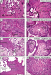
Histopathological changes in untreated HCMV-infected placentas. A and B: Fibrotic villi with necrosis, calcification, and hemosiderin deposits. C and D: Fibrin-encased avascular villi and fibrosis in the villus core. E and F: Area of fibrin deposition surrounded by severely hydropic villi. G and H: Chorionic villitis with plasma cell infiltration. H&E stain. Original magnification: ×100 (A–H); ×400 (insets). BV, blood vessel.
Figure 3
Figure 3
Significant increase in the number of chorionic villi in placentas in the HIG treatment group. Cross-sections of paraffin-embedded placentas were immunostained for trophoblasts with anti-cytokeratin-7 antibody. Nuclei were counterstained with hematoxylin. Original magnification, ×200. The number of villi per mm2 was counted (three to four placentas/group, 100 to 200 microscopic fields; original magnification ×200). Representative placentas are from control (A), untreated congenital HCMV infection (B), and HIG treatment (C) groups. D: Graph showing the ratios of average counts relative to controls using Poisson regression.
Figure 4
Figure 4
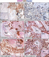
Significant increase in the number of villous blood vessels in congenitally infected placentas. Cross-sections of paraffin-embedded placental biopsy specimens were immunostained for blood vessels with anti-CD34 (brown) and anti-CD31 (fuchsia) antibodies. Nuclei were counterstained with hematoxylin. Original magnification, ×100. The number of blood vessels per villus was counted (3 to 4 placentas/group, 100 to 200 microscopic fields; insets original magnification, ×400). Representative placentas are from uninfected control (A), untreated congenital infection (B and C), and HIG treatment (D) groups. E: Graph showing the ratios of average blood vessel counts relative to controls using Poisson regression.
Figure 5
Figure 5
Up-regulated VEGF expression in congenitally infected placentas. Cross-sections of paraffin-embedded placental biopsy specimens were immunostained for VEGF (fuchsia) and cytokeratin 7 (brown). Nuclei were counterstained with hematoxylin. Original magnification: ×100 (A–F); ×400 (insets). Representative decidua (A, C, and E) and placentas (B, D, and F) were from uninfected control (A and B), untreated infection (C and D), and HIG treatment (E and F) groups. BV, blood vessel; CTB, cytotrophoblasts; ST, syncytiotrophoblasts; Mφ, macrophages.
Figure 6
Figure 6
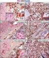
Flt1 expression in congenitally infected placentas. Cross-sections of paraffin-embedded placental biopsy specimens were immunostained for Flt1 (fuchsia) and cytokeratin 7 (brown). Nuclei were counterstained with hematoxylin. Original magnification: ×100 (A–F); ×400 (insets). Representative decidua (A, C, and E) and placentas (B, D, and F) were from control (A and B), untreated infection (C and D), and HIG treatment (E and F) groups. BV, blood vessel; CTB, cytotrophoblasts; ST, syncytiotrophoblasts; Mφ, macrophages.
Figure 7
Figure 7
Enhanced VEGF and Flt1 expression in placentas with congenital CMV infection. Levels of VEGF (A) and Flt1 (B) expression were quantified by immunostaining paraffin-embedded placentas from uninfected control (n = 3), untreated HCMV infection (n = 4), and HIG treatment (n = 4) groups. Intensities of DAB-labeled single immunostainings were evaluated using a semiquantitative digital imaging method.,, The number of digitally quantified counts was 15 for uninfected control, 23 for untreated HCMV infection, and 15 for HIG treatment placentas. Staining intensity was expressed as the mean staining intensity score ± SEM (bars) and compared between groups using Bonferroni correction of P values for multiple comparisons and Bootstrap resampling using 1000 replications.
Figure 8
Figure 8
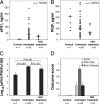
Increase in sFlt1/PlGF ratios in amniotic fluid from women with untreated congenital HCMV infection associated with poor outcome score. Values of sFlt1 (A), PlGF (B), and the mean sFlt1/PlGF ratio after logarithmic transformation (C) in uninfected control (n = 7), untreated congenital infection (n = 38), and HIG treatment (n = 9) groups. A and B: Median values of each set of data points are indicated by a horizontal bar C: Results are expressed as mean ± SEM and compared between groups using Bonferroni correction of P values for multiple comparisons. Differences were significant between untreated infection and control groups (P < 0.001) and between HIG treatment and control groups (P < 0.001) but not between untreated infection and HIG treatment groups (P = 0.135). D: Fetal outcomes in control, untreated infection, and HIG treatment groups were scored as 0 for no symptoms and 1 for symptoms (IUGR, HCMV DNA in urine at birth, or brain disease). The outcome score is expressed as mean ± SEM.

References

    1. Kenneson A, Cannon MJ. Review and meta-analysis of the epidemiology of congenital cytomegalovirus (CMV) infection. Rev Med Virol. 2007;17:253–276. - PubMed
    1. Colugnati FA, Staras SA, Dollard SC, Cannon MJ. Incidence of cytomegalovirus infection among the general population and pregnant women in the United States. BMC Infect Dis. 2007;7:71. - PMC - PubMed
    1. Cannon MJ, Pellett PE. Risk of congenital cytomegalovirus infection. Clin Infect Dis. 2005;40:1701–1702. author reply 1702–1703. - PubMed
    1. Demmler GJ. Congenital cytomegalovirus infection and disease. Adv Pediatr Infect Dis. 1996;11:135–162. - PubMed
    1. Pass RF, Fowler KB, Boppana SB, Britt WJ, Stagno S. Congenital cytomegalovirus infection following first trimester maternal infection: symptoms at birth and outcome. J Clin Virol. 2006:216–220. - PubMed

Publication types

MeSH terms

Substances