. 2010 Sep;177(3):1065-71.
doi: 10.2353/ajpath.2010.090923. Epub 2010 Jul 22.

Resveratrol inhibits renal fibrosis in the obstructed kidney: potential role in deacetylation of Smad3

Affiliations

Resveratrol inhibits renal fibrosis in the obstructed kidney: potential role in deacetylation of Smad3

Jinhua Li et al. Am J Pathol. 2010 Sep.

Abstract

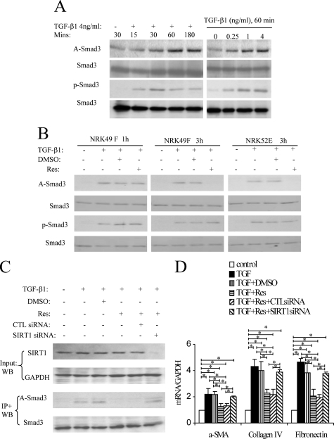
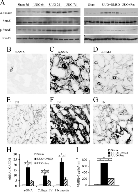
Transforming growth factor-beta1 (TGF-beta1) promotes tissue fibrosis through the Smad3 signaling pathway. While phosphorylation is known to regulate Smad3 function, recent in vitro studies have suggested that acetylation may also regulate Smad3 function. This study investigated Smad3 acetylation in renal fibrosis. TGF-beta1 stimulation of renal fibroblasts and tubular epithelial cells induced Smad3 acetylation and phosphorylation. Resveratrol, an activator of the Nicotinamide adenine dinucleotide (NAD) dependent protein deacetylase SIRT1, reversed acetylation but not phosphorylation of Smad3 and inhibited TGF-beta1-induced up-regulation of collagen IV and fibronectin mRNA levels. Knockdown of SIRT1 expression abolished the inhibitory effect of resveratrol, and co-immunoprecipitation studies provide direct evidence of an interaction between acetylated Smad3 and SIRT1. The role of Smad3 acetylation in renal fibrosis was then examined in the unilateral ureteric obstruction (UUO) model. Immunoprecipitation studies showed acetylation and phosphorylation of Smad3 by day 2 UUO, which was sustained to day 7 in association with development of interstitial fibrosis. Resveratrol inhibited acetylation but not phosphorylation of Smad3 at day 2 UUO, and resveratrol treatment inhibited interstitial fibrosis at day 7 UUO. In conclusion, these studies support a pathological role for Smad3 acetylation in renal fibrosis and suggest that deacetylation of Smad3 may be a novel therapeutic target for fibrotic disease.


Figures

Figure 1
Figure 1
TGF-β1-induced Smad3 acetylation (A-Smad3) and phosphorylation (p-Smad3) in rat renal NRK49F fibroblasts. A: Immunoprecipitation (IP)/Western blotting (WB) shows a time course of TGF-β1 (4 ng/ml)-induced Smad3 acetylation and phosphorylation, and a TGF-β1 dose-response at the 60-minute time point. B: IP/WB analysis of the effect of Resveratrol (Res) on TGF-β1-induced Smad3 acetylation and phosphorylation. The addition of 10 μmol/L Res to NRK49F cells suppressed Smad3 acetylation at three hours but not one hour, while the addition of 5 μmol/L Res suppressed Smad3 acetylation at 3 hours in NRK52E cells. C: WB in top panel shows that SIRT1 siRNA but not control siRNA knocks down SIRT1 protein levels in NRK49F cells. IP/WB analysis in bottom panel shows that TGF-β1-induced Smad3 acetylation at three hours is suppressed by Res, but this suppression is lost in the presence of SIRT1 siRNA. D: Real-time RT-PCR analysis of mRNA levels in NRK49F cells 24 hours after TGF-β1 stimulation. The addition of 10 μmol/L Res suppressed TGF-β1-induced up-regulation of α-SMA, collagen IV, and fibronectin, and this effect of Res was prevented by SIRT1 siRNA but not control siRNA. Data are mean ± SD. *P < 0.05.
Figure 2
Figure 2
Evidence of direct interaction of Smad3 and SIRT1 in TGF-β1–stimulated NRK49F cells. A: Smad3 immunoprecipitation (IP) from TGF-β1 (4 ng/ml for 1, 3 or 6 hours) stimulated cells was examined by Western blotting (WB) showing Smad3 acetylation (α-A-lysine), co-immunoprecipitation of Smad3 and p300, and of Smad3 with SIRT1. B: NRK49F cells were cotransfected with plasmids expressing Smad3 and Flag-SIRT1. IP/WB showed co-immunoprecipitation of Smad3 and SIRT1 at three hours and six hours after TGF-β1 stimulation. C: NRK49F cells were transfected with a Smad-binding element (SBE)-luciferase reporter construct and a control reporter (PRL-TK) to measure Smad transcriptional activity. After 18 hours, cells were stimulated with TGF-β1 for a further 18 hours in the presence of 10 μmol/L Resveratrol (Res), DMSO vehicle, or SIRT1 activator three and then cells harvested and dual-luciferase activity assayed. Data are mean ± SD. *P < 0.05; ***P < 0.001. D: NRK49F cells were transfected with the SBE and control reporters and with plasmids expressing Smad3, SIRT1, or SIRT1 H363Y (dominant negative form). After 18 hours, cells were stimulated with TGF-β1 for a further 18 hours and then cells harvested and dual-luciferase activity assayed. Data are mean ± SD. *P < 0.05; ***P < 0.001. E and F: NRK49F cells were transfected with control siRNA, Smad2 siRNA, and Smad3 siRNA. After 48 hours, cells were harvested for western blotting (E) or stimulated with TGF-β1 for a further 24 hours and then cells harvested and real-time PCR was performed to detect α-SMA. Data are mean ± SD. ***P < 0.001.
Figure 3
Figure 3
Smad3 acetylation following unilateral ureter obstruction (UUO). A: Left panel shows immunoprecipitation/Western blotting of Smad3 acetylation (A-Smad3) and phosphorylation (p-Smad3) in a time course of UUO or sham-operated mice. Right panel shows that Resveratrol (Res) treatment prevented Smad3 acetylation, but not Smad3 phosphorylation, on day 2 UUO. Immunofluorescence staining at day 7 UUO showing α-smooth muscle actin (α-SMA) in sham-operated (B), UUO+Vehicle (C), and UUO+Res (D); and immunofluorescence staining for fibronectin in sham operated (E), UUO+Vehicle (F), and UUO+Res (G). H: Real-time RT-PCR analysis of kidney α-SMA, collagen IV, and fibronectin mRNA levels at day seven UUO. I: Quantification of immunofluorescence staining for F4/80+ macrophages at day seven UUO. Data are mean ± SD. *P < 0.05.

References

    1. Flanders KC. Smad3 as a mediator of the fibrotic response. Int J Exp Pathol. 2004;85:47–64. - PMC - PubMed
    1. Wang W, Koka V, Lan HY. Transforming growth factor-beta and Smad signalling in kidney diseases. Nephrology. 2005;10:48–56. - PubMed
    1. Sato M, Muragaki Y, Saika S, Roberts AB, Ooshima A. Targeted disruption of TGF-β1/Smad3 signaling protects against renal tubulointerstitial fibrosis induced by unilateral ureteral obstruction. J Clin Invest. 2003;112:1486–1494. - PMC - PubMed
    1. Fujimoto M, Maezawa Y, Yokote K, Joh K, Kobayashi K, Kawamura H, Nishimura M, Roberts AB, Saito Y, Mori S. Mice lacking Smad3 are protected against streptozotocin-induced diabetic glomerulopathy. Biochem Biophys Res Commun. 2003;305:1002–1007. - PubMed
    1. Li JH, Huang XR, Zhu HJ, Oldfield M, Cooper M, Truong LD, Johnson RJ, Lan HY. Advanced glycation end products activate Smad signaling via TGF-beta-dependent and independent mechanisms: implications for diabetic renal and vascular disease. FASEB J. 2004;18:176–178. - PubMed

Publication types

MeSH terms