. 2010 Aug 10;107(32):14339-44.
doi: 10.1073/pnas.0912701107. Epub 2010 Jul 22.

Identification of a microRNA signature of renal ischemia reperfusion injury

Affiliations

Identification of a microRNA signature of renal ischemia reperfusion injury

Jonathan G Godwin et al. Proc Natl Acad Sci U S A. .

Abstract

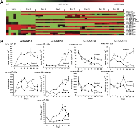
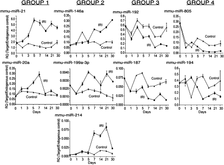
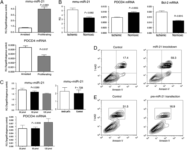
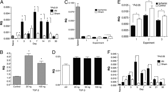
Renal ischemia reperfusion injury (IRI) is associated with significant morbidity and mortality. Given the importance of microRNAs (miRNAs) in regulating gene expression, we examined expression profiles of miRNAs following renal IRI. Global miRNA expression profiling on samples prepared from the kidneys of C57BL/6 mice that underwent unilateral warm ischemia revealed nine miRNAs (miR-21, miR-20a, miR-146a, miR-199a-3p, miR-214, miR-192, miR-187, miR-805, and miR-194) that are differentially expressed following IRI when compared with sham controls. These miRNAs were also differently expressed following IRI in immunodeficient RAG-2/common gamma-chain double-knockout mice, suggesting that the changes in expression observed are not significantly influenced by lymphocyte infiltration and therefore define a lymphocyte-independent signature of renal IRI. In vitro studies revealed that miR-21 is expressed in proliferating tubular epithelial cells (TEC) and up-regulated by both cell-intrinsic and -extrinsic mechanisms resulting from ischemia and TGF-beta signaling, respectively. In vitro, knockdown of miR-21 in TEC resulted in increased cell death, whereas overexpression prevented cell death. However, overexpression of miR-21 alone was not sufficient to prevent TEC death following ischemia. Our findings therefore define a molecular fingerprint of renal injury and suggest miR-21 may play a role in protecting TEC from death.


Conflict of interest statement

The authors declare no conflict of interest.

Figures

Fig. 1.
Fig. 1.
Identification of a miRNA signature following renal IRI. (A) Heat map of miRNAs differentially expressed in the kidneys of C57BL/6J mice after IRI or sham operation. Mean signal intensities (n = 3) following normalization and z-transformation. (B) Plots of above microarray data expressed as the mean signal intensity ± SE. Data were normalized using a locally weighted regression method. Naïve kidneys serve as day 0. Data grouped based upon similar expression patterns over time.
Fig. 2.
Fig. 2.
The miRNA fingerprint is lymphocyte independent. The expression of the indicated miRNAs was measured in Ro/cγ0 mice by real-time PCR. The data are expressed as mean ± SE of three independent experiments. Relative quantitation (RQ) was calculated by the ddCT method normalizing miRNA expression to the endogenous control SnoRNA202.
Fig. 3.
Fig. 3.
miR-21 regulates proliferation and apoptosis in TEC. (A) Real-time PCR analysis of miR-21 and PDCD4 in arrested and proliferating primary TEC cultures. Cells were growth arrested by growing to confluence. The relative expression of miR-21 was compared in growth-arrested and proliferating cultures by real-time PCR. (B) Expression of miR-21, PDCD4, and Bcl-2 in primary TEC following ischemia. The relative expression of miR-21, PDCD4, and Bcl-2 were compared in ischemic and normoxic TEC cultures at 48 h. (C Left) Knockdown of miR-21 in primary TEC cultures. TEC cultures were transfected with increasing concentrations of anti-miR-21 LNA oligonucleotide and analyzed 48 h later. (Right) Knockdown of an irrelevant miRNA, let-7c, fails to influence miR-21 expression and analyzed 48 h later. TEC were transfected with anti-let-7c LNA oligonucleotides. The levels of miR-21 and PDCD4 were quantitated by real-time PCR. Data in A–C are expressed as relative quantifications (RQ) by normalizing to the endogenous control SnoRNA202 (miR-21) or GAPDH (PDCD4, Bcl-2). (D) Induction of apoptosis in TEC following miR-21 knockdown. Twenty-four hours after transfection with miR-21 or Fugene alone (control), cell death was analyzed by staining for Annexin-V and 7-AAD and quantified by flow cytometry. The percent of dead cells is indicated. (E) Overexpression of miR-21 results in decreased TEC death. Twenty-four hours after transfection with premiR-21 or Fugene alone (control), cell death was analyzed by staining for Annexin-V and 7-AAD and quantified by flow cytometry as above. In all cases, the data shown are representative of at least two experiments.
Fig. 4.
Fig. 4.
TGF-β-induced miR-21 expression in TEC. (A) Analysis of TGF-β expression in the kidneys of C57BL/6 mice undergoing IRI or sham surgery. Kidneys from naïve C57BL/6 mice were used as a control. Shown is expression on various days after injury or sham surgery. TGF-β expression was analyzed by real-time PCR. Pooled RNA from at least three kidneys per time point was analyzed. (B) Cultures of primary TEC were established from C57BL/6 mice and stimulated with either 20 or 100 ng/mL TGF-β for 24 h. The cells were harvested and total RNA prepared. The levels of mature miR-21 were then determined by real-time PCR. Similar results were observed at 72 h. Shown are data from at least three separate experiments. Relative quantitation (RQ) was calculated by the ddCT method normalizing miRNA expression to the endogenous control SnoRNA202. (C) TEC do not produce TGF-β following ischemia. Cultures of primary TEC were established as above and ischemia induced. Sham cells were not overlaid with mineral oil. The cells were harvested and TGF-β expression analyzed by RT-PCR using standard methods. Spleen cells were used as a positive control. Shown are data from at least three separate experiments. RQ was calculated by normalizing expression to the endogenous control GAPDH. (D) TGF-β stimulation leads to a modest increase in pri-miR-21. Cultures of primary TEC were established and stimulated with the indicated amounts of TGF-β for 24 h. The cells were harvested and total RNA prepared. Levels of pri-miR-21 were then quantitated by real-time PCR using the ABI TaqMan mmu-pri-miR-21 assay kit. (E) Ischemia leads to a decrease in pri-miR-21. Cultures of primary TEC were established, ischemia was induced, and the cells were harvested. Levels of pri-miR-21 were then quantitated as indicated for C. (F) Analysis of pri-miR-21 expression in the kidneys of C57BL/6 mice undergoing IRI or sham surgery. Kidneys from naïve C57BL/6 mice were used as a control. Shown is expression on various days after injury or sham surgery. Pooled RNA from at least three kidneys per time point was analyzed.

References

    1. Thadhani R, Pascual M, Bonventre JV. Acute renal failure. N Engl J Med. 1996;334:1448–1460. - PubMed
    1. Chertow GM, Burdick E, Honour M, Bonventre JV, Bates DW. Acute kidney injury, mortality, length of stay, and costs in hospitalized patients. J Am Soc Nephrol. 2005;16:3365–3370. - PubMed
    1. Bonventre JV, Zuk A. Ischemic acute renal failure: An inflammatory disease? Kidney Int. 2004;66:480–485. - PubMed
    1. Koo DD, Welsh KI, Roake JA, Morris PJ, Fuggle SV. Ischemia/reperfusion injury in human kidney transplantation: An immunohistochemical analysis of changes after reperfusion. Am J Pathol. 1998;153:557–566. - PMC - PubMed
    1. Jang HR, Ko GJ, Wasowska BA, Rabb H. The interaction between ischemia-reperfusion and immune responses in the kidney. J Mol Med. 2009;87:859–864. - PubMed

Publication types

LinkOut - more resources