. 2010 Oct;30(10):1940-8.
doi: 10.1161/ATVBAHA.110.205997. Epub 2010 Jul 22.

Acute humanin therapy attenuates myocardial ischemia and reperfusion injury in mice

Affiliations

Acute humanin therapy attenuates myocardial ischemia and reperfusion injury in mice

Radhika H Muzumdar et al. Arterioscler Thromb Vasc Biol. 2010 Oct.

Abstract

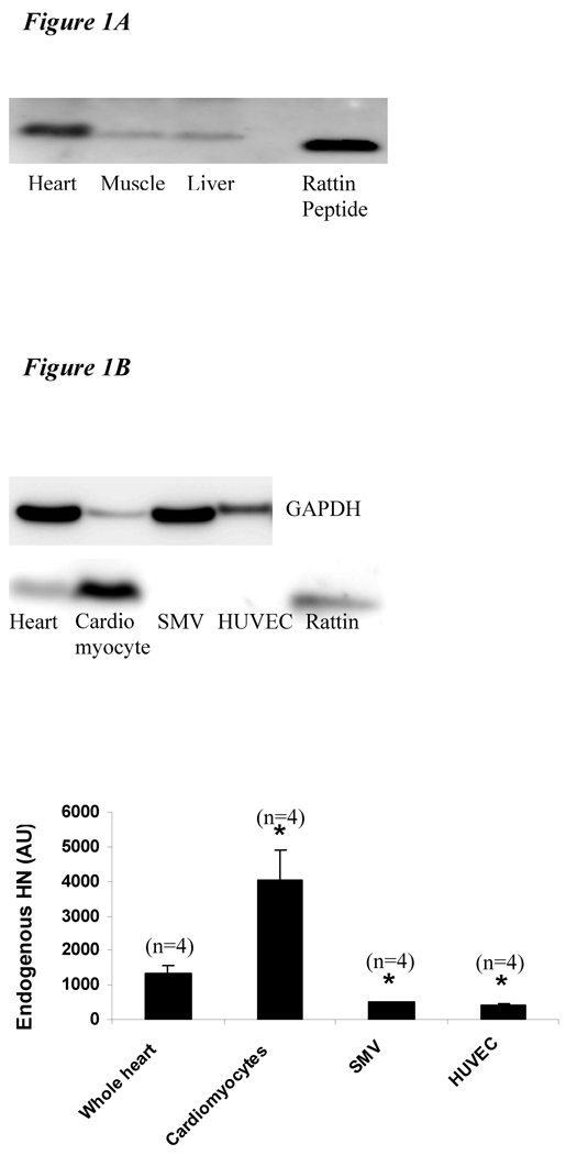
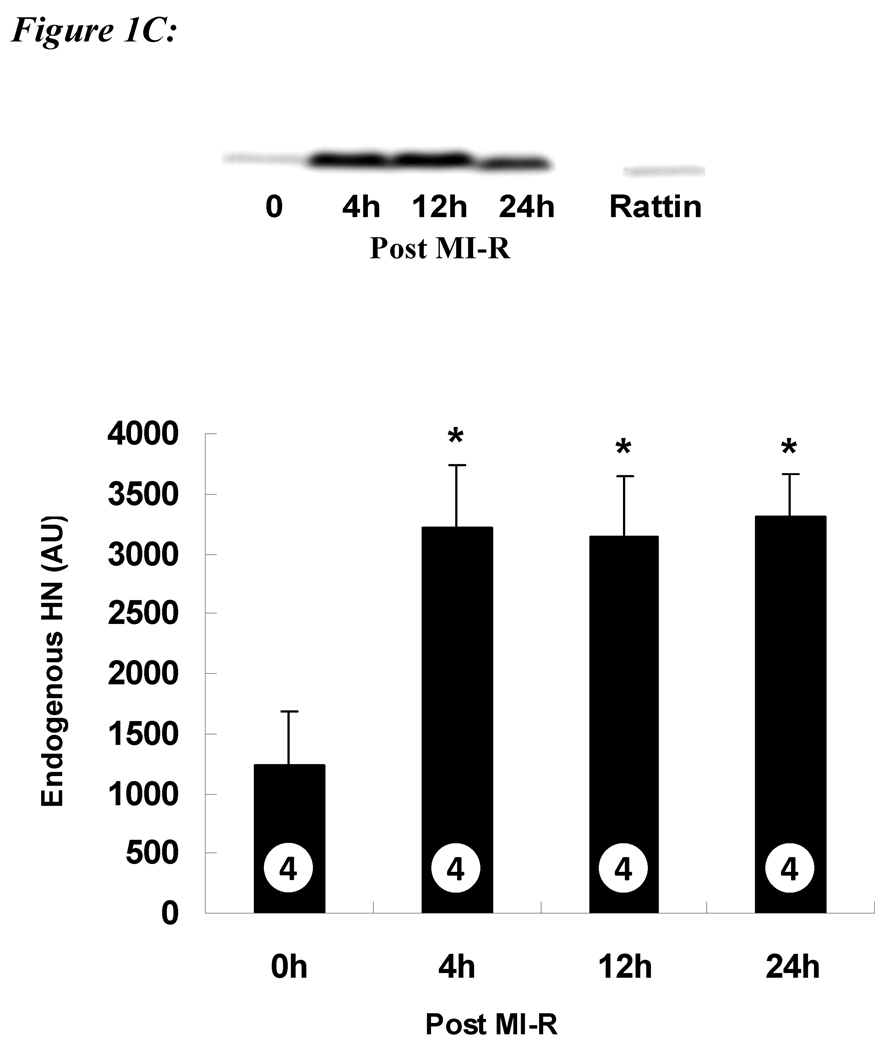
Objective: Humanin (HN), an endogenous antiapoptotic peptide, has previously been shown to protect against Alzheimer's disease and a variety of cellular insults. We evaluated the effects of a potent analog of HN (HNG) in an in vivo murine model of myocardial ischemia and reperfusion.

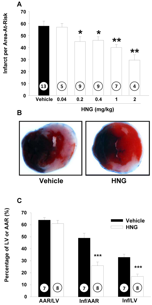
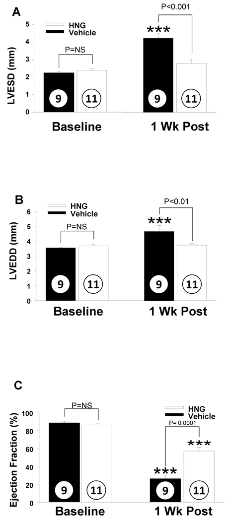
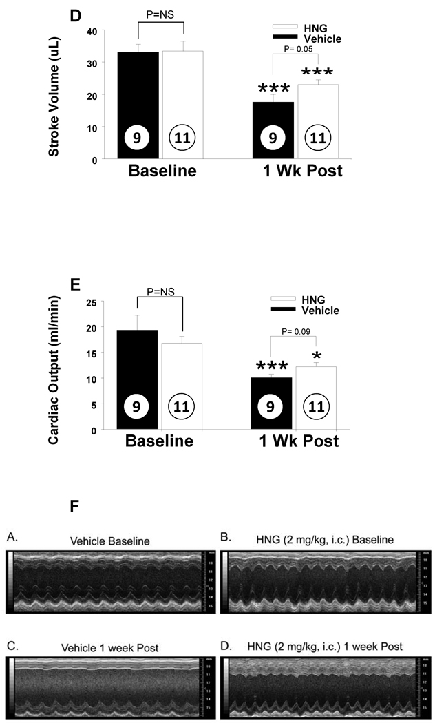
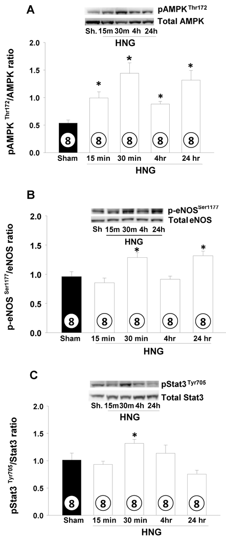
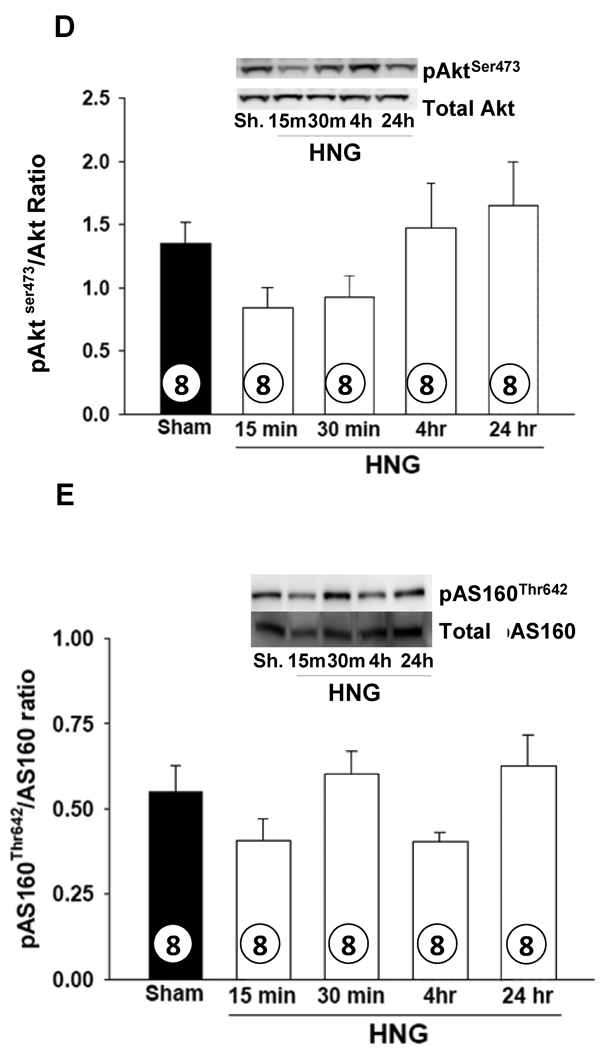
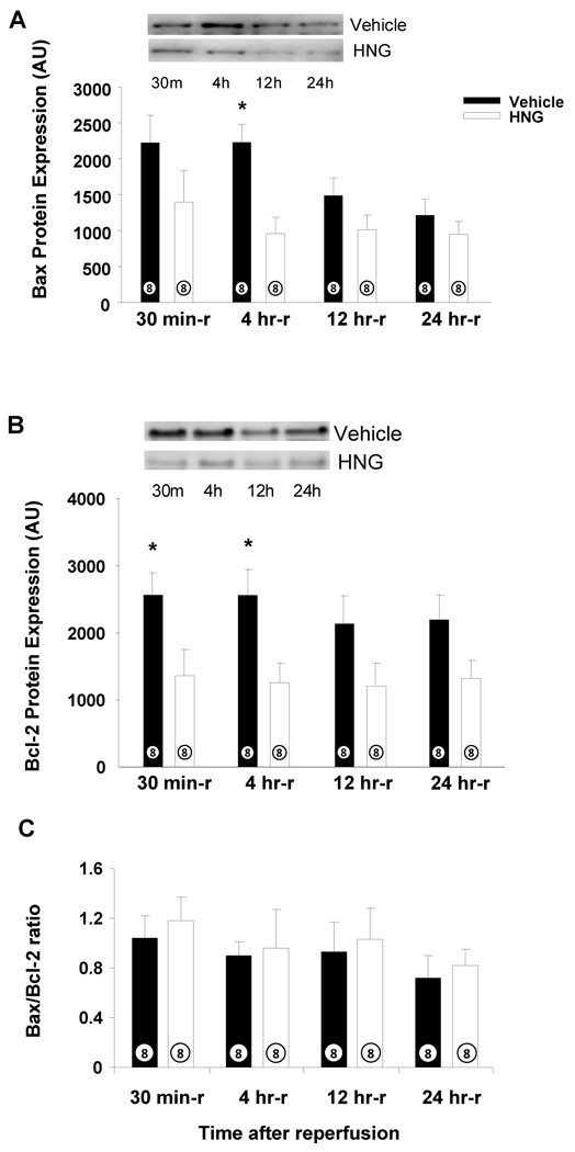
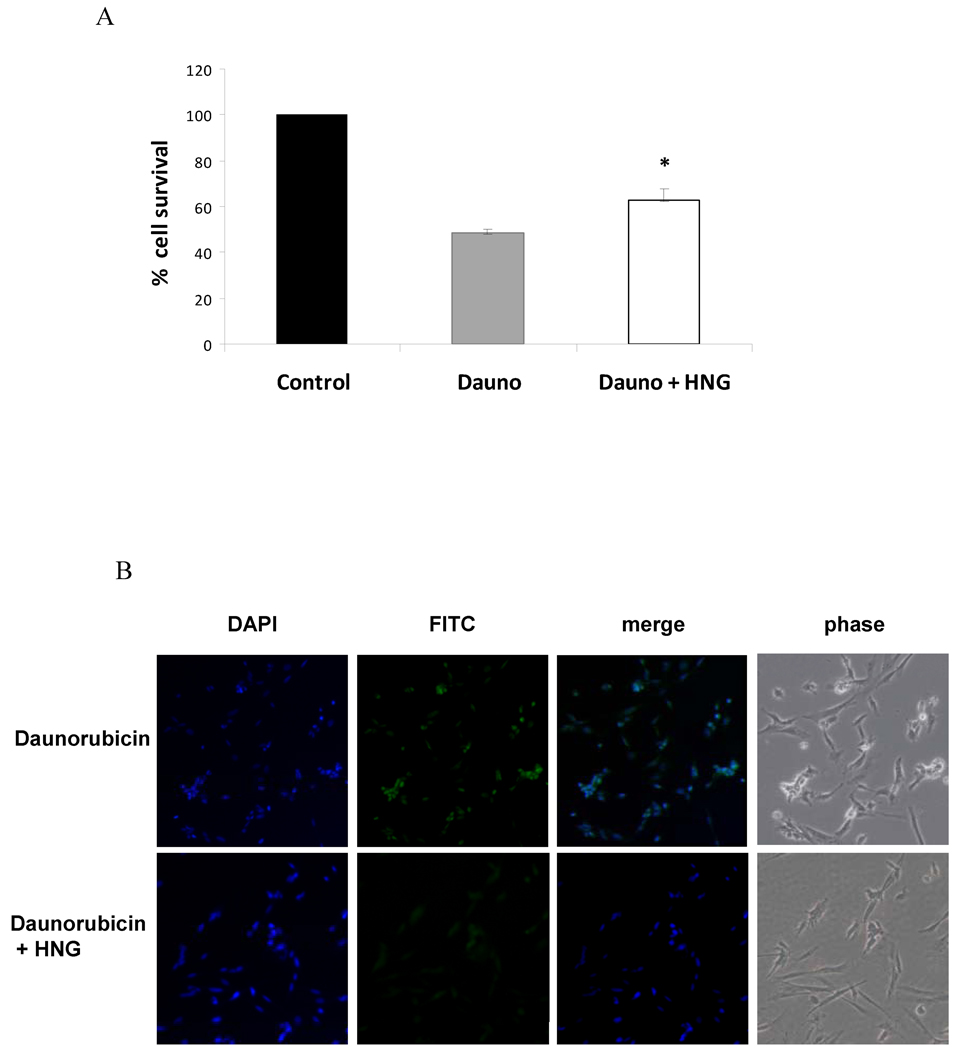
Methods and results: Male C57BL6/J mice (8 to 10 week old) were subjected to 45 minutes of left coronary artery occlusion followed by a 24-hour reperfusion. HNG or vehicle was administered IP 1 hour prior or at the time of reperfusion. The extent of myocardial infarction per area-at-risk was evaluated at 24 hours using Evans Blue dye and 2-3-5-triphenyl tetrazolium chloride staining. Left ventricular function was evaluated at 1 week after ischemia using high-resolution, 2D echocardiography (VisualSonics Vevo 770). Myocardial cell signaling pathways and apoptotic markers were assessed at various time points (0 to 24 hours) following reperfusion. Cardiomyocyte survival and apoptosis in response to HNG were assessed in vitro. HNG reduced infarct size relative to the area-at-risk in a dose-dependent fashion, with a maximal reduction at the dose of 2 mg/kg. HNG therapy enhanced left ventricular ejection fraction and preserved postischemic left ventricular dimensions (end-diastolic and end-systolic), resulting in improved cardiac function. Treatment with HNG significantly increased phosphorylation of AMPK and phosphorylation of endothelial nitric oxide synthase in the heart and attenuated Bcl-2-associated X protein and B-cell lymphoma-2 levels following myocardial ischemia and reperfusion. HNG improved cardiomyocyte survival and decreased apoptosis in response to daunorubicin in vitro.

Conclusions: These data show that HNG provides cardioprotection in a mouse model of myocardial ischemia and reperfusion potentially through activation of AMPK-endothelial nitric oxide synthase-mediated signaling and regulation of apoptotic factors. HNG may represent a novel agent for the treatment of acute myocardial infarction.


Figures

Figure 1
Figure 1
A) Protein level of HN in different tissues in mice: A total of 30 µg of protein from mouse heart (lane 1), skeletal muscle (lane 2), liver (lane 3) and 3 ng of synthetic RN (rodent homolog of HN) peptide (lane 5) were loaded. Higher amounts of HN protein were detected in heart compared to muscle and liver. B) Protein level of HN in different cell types in heart: 30 µg of mouse heart (lane 1), cardiomyocyte (lane 2), smooth muscle vascular cell (lane 3), human umbilical vein endothelial cells (lane 4) and 3 ng of synthetic RN peptide (lane 5) were loaded. HN levels were highest in the cardiomyocytes (*p< 0.001 vs. other cell lines). C) Protein levels of HN post MI-R: Protein extracts from left ventricle at basal (lane 1), 4 hr (lane 2), 12 hr (lane 3) and 24 hr (lane 4) post MI-R were loaded. The levels of HN increase post MI-R and persist up to 24 hrs post injury (*p < 0.01 vs. baseline).
Figure 1
Figure 1
A) Protein level of HN in different tissues in mice: A total of 30 µg of protein from mouse heart (lane 1), skeletal muscle (lane 2), liver (lane 3) and 3 ng of synthetic RN (rodent homolog of HN) peptide (lane 5) were loaded. Higher amounts of HN protein were detected in heart compared to muscle and liver. B) Protein level of HN in different cell types in heart: 30 µg of mouse heart (lane 1), cardiomyocyte (lane 2), smooth muscle vascular cell (lane 3), human umbilical vein endothelial cells (lane 4) and 3 ng of synthetic RN peptide (lane 5) were loaded. HN levels were highest in the cardiomyocytes (*p< 0.001 vs. other cell lines). C) Protein levels of HN post MI-R: Protein extracts from left ventricle at basal (lane 1), 4 hr (lane 2), 12 hr (lane 3) and 24 hr (lane 4) post MI-R were loaded. The levels of HN increase post MI-R and persist up to 24 hrs post injury (*p < 0.01 vs. baseline).
Figure 2
Figure 2
HNG decreases myocardial infarct size in mice following MI-R: A) myocardial infarct size in mice receiving doses of HNG ranging from 0.2 to 2 mg/kg. HNG significantly decreased myocardial infarct size compared with vehicle in a dose dependent manner. B) Representative mid-ventricular photomicrographs of mouse hearts are shown after 45 min of myocardial ischemia and 24 hr of reperfusion. Areas of the myocardium that appear blue represent the areas of myocardium not at risk for infarction. In contrast, the areas of myocardium that stain red (i.e., TTC positive) represent viable myocardium that was at risk for infarction. Myocardium that appears pale (i.e., TTC negative) indicates areas of myocardium at risk that are necrotic (i.e., infarcted). C) HNG administered at the time of reperfusion decreased infarct size significantly compared to AAR. Values are means ± SE. Numbers inside bars indicate the number of animals investigated in each group. **p <0.01; ***p <0.001 vs. vehicle.
Figure 3
Figure 3
HNG treatment improves cardiac function post MI-R: Cardiac function post MIR was evaluated by Echocardiography. Mice treated with HNG had significantly improved cardiac function as evidenced by effects on A) LVESD B) LVEDD C) Ejection fraction D) Stroke Volume and E) Cardiac Output. F) Representative baseline and post MIR M-mode images of the LV from vehicle and HNG treated mice. Values are means ± SE. **p <0.01; ***p <0.001 vs. baseline.
Figure 3
Figure 3
HNG treatment improves cardiac function post MI-R: Cardiac function post MIR was evaluated by Echocardiography. Mice treated with HNG had significantly improved cardiac function as evidenced by effects on A) LVESD B) LVEDD C) Ejection fraction D) Stroke Volume and E) Cardiac Output. F) Representative baseline and post MIR M-mode images of the LV from vehicle and HNG treated mice. Values are means ± SE. **p <0.01; ***p <0.001 vs. baseline.
Figure 4
Figure 4
HNG activates cardiac AMPK and eNOS signaling pathways.Effects of HNG or vehicle over a time course on A) pAMPKThr172 B) phospho-eNOSSer1177 C) pSTAT-3Tyr705 D) pAktS473 and E) pAS160Thr642 levels. Values are means ± SE. * p< 0.05, significantly different from SHAM.
Figure 4
Figure 4
HNG activates cardiac AMPK and eNOS signaling pathways.Effects of HNG or vehicle over a time course on A) pAMPKThr172 B) phospho-eNOSSer1177 C) pSTAT-3Tyr705 D) pAktS473 and E) pAS160Thr642 levels. Values are means ± SE. * p< 0.05, significantly different from SHAM.
Figure 5
Figure 5
HNG down-regulates Bax following MI-R: Effects of HNG or vehicle on A) Bax levels post MI-R. B) Bcl-2 levels post MI-R. C) Bax/Bcl-2 ratio post MI-R. Values are means ± SE. * p < 0.05, significantly different from vehicle.
Figure 6
Figure 6
HNG improves cardiomyocyte survival in vitro and decreases apoptosis in response to Daunorubicin. Effect of HNG on A) Cardiomyocyte survival as assessed by cell viability assay, * p < 0.05 Dauno compared to Dauno+HNG, and B) early markers of apoptosis as assessed by CaspASE FITC -VAD-FMK in situ apoptosis assay.

References

    1. Kung HC, Hoyert DL, Xu J, Murphy SL. Deaths: final data for 2005. Natl Vital Stat Rep. 2008;56:1–120. - PubMed
    1. Lloyd-Jones D, Adams R, Carnethon M, De Simone G, Ferguson TB, Flegal K, Ford E, Furie K, Go A, Greenlund K, Haase N, Hailpern S, Ho M, Howard V, Kissela B, Kittner S, Lackland D, Lisabeth L, Marelli A, McDermott M, Meigs J, Mozaffarian D, Nichol G, O'Donnell C, Roger V, Rosamond W, Sacco R, Sorlie P, Stafford R, Steinberger J, Thom T, Wasserthiel-Smoller S, Wong N, Wylie-Rosett J, Hong Y. Heart disease and stroke statistics--2009 update: a report from the American Heart Association Statistics Committee and Stroke Statistics Subcommittee. Circulation. 2009;119:480–486. - PubMed
    1. Muzumdar RH, Huffman DM, Atzmon G, Buettner C, Cobb LJ, Fishman S, Budagov T, Cui L, Einstein FH, Poduval A, Hwang D, Barzilai N, Cohen P. Humanin: a novel central regulator of peripheral insulin action. PLoS One. 2009;4:e6334. - PMC - PubMed
    1. Jung SS, Van Nostrand WE. Humanin rescues human cerebrovascular smooth muscle cells from Abeta-induced toxicity. J Neurochem. 2003;84:266–272. - PubMed
    1. Hashimoto Y, Niikura T, Tajima H, Yasukawa T, Sudo H, Ito Y, Kita Y, Kawasumi M, Kouyama K, Doyu M, Sobue G, Koide T, Tsuji S, Lang J, Kurokawa K, Nishimoto I. A rescue factor abolishing neuronal cell death by a wide spectrum of familial Alzheimer's disease genes and Abeta. Proc Natl Acad Sci U S A. 2001;98:6336–6341. - PMC - PubMed

Publication types

MeSH terms

LinkOut - more resources