Comparative Study
. 2010 Sep 17;107(6):737-46.
doi: 10.1161/CIRCRESAHA.109.215715. Epub 2010 Jul 22.

Identification of a novel macrophage phenotype that develops in response to atherogenic phospholipids via Nrf2

Affiliations
Comparative Study

Identification of a novel macrophage phenotype that develops in response to atherogenic phospholipids via Nrf2

Alexandra Kadl et al. Circ Res. .

Abstract

Rationale: Macrophages change their phenotype and biological functions depending on the microenvironment. In atherosclerosis, oxidative tissue damage accompanies chronic inflammation; however, macrophage phenotypic changes in response to oxidatively modified molecules are not known.

Objective: To examine macrophage phenotypic changes in response to oxidized phospholipids that are present in atherosclerotic lesions.

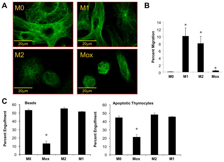
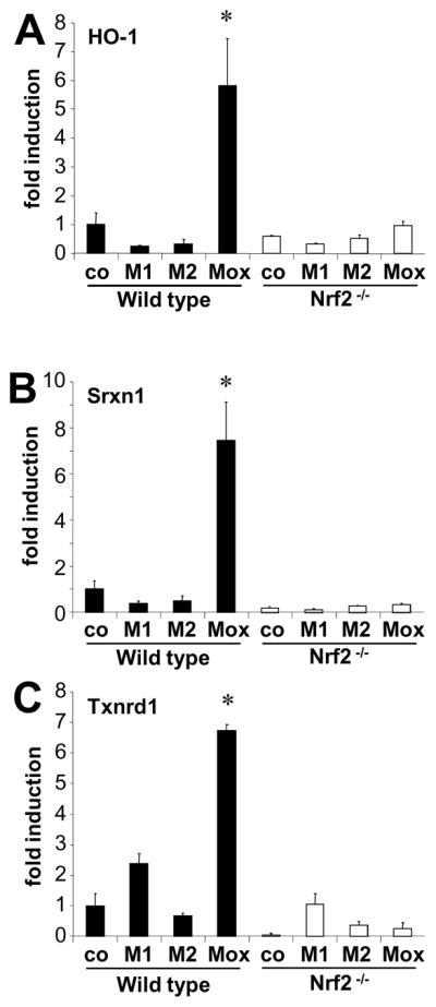
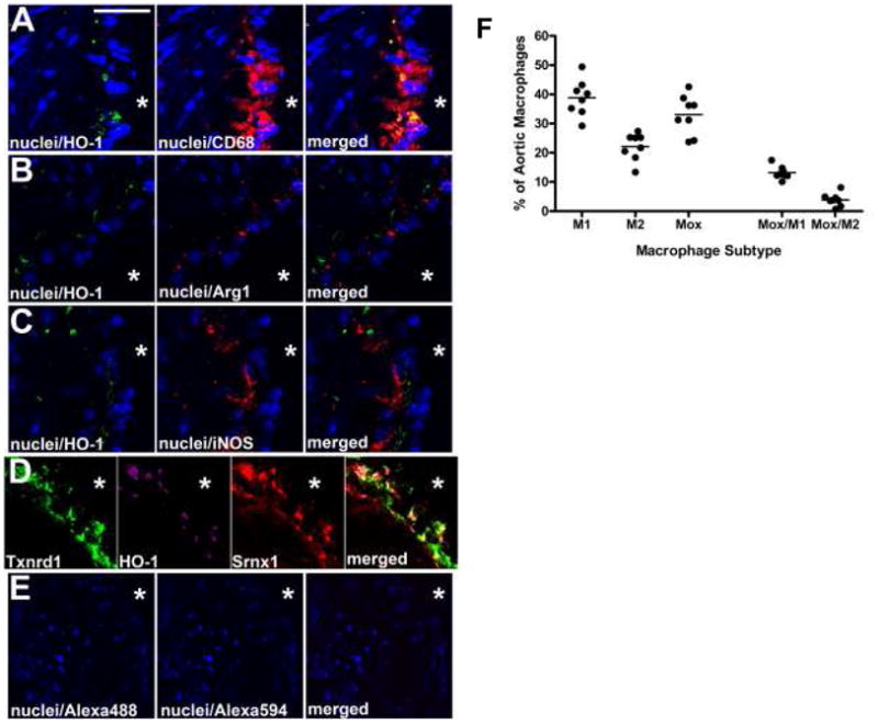
Methods and results: We show that oxidized phospholipid-treated murine macrophages develop into a novel phenotype (Mox) that is strikingly different from the conventional M1 and M2 macrophage phenotypes. Compared to M1 and M2, Mox macrophages show a different gene expression pattern, as well as decreased phagocytotic and chemotactic capacity. Treatment with oxidized phospholipids induces both M1 and M2 macrophages to switch to the Mox phenotype. Whole-genome expression array analysis and subsequent gene ontology clustering revealed that the Mox phenotype was characterized by abundant overrepresentation of Nrf2-mediated expression of redox-regulatory genes. In macrophages isolated from Nrf2(-/-) mice, oxidized phospholipid-induced gene expression and regulation of redox status were compromised. Moreover, we found that Mox macrophages comprise 30% of all macrophages in advanced atherosclerotic lesions of low-density lipoprotein receptor knockout (LDLR(-/-)) mice.

Conclusions: Together, we identify Nrf2 as a key regulator in the formation of a novel macrophage phenotype (Mox) that develops in response to oxidative tissue damage. The unique biological properties of Mox macrophages suggest this phenotype may play an important role in atherosclerotic lesion development as well as in other settings of chronic inflammation.


Figures

Figure 1
Figure 1. Mox macrophages represent a functionally distinct phenotype
Bone marrow-derived macrophages were polarized with either 10U/ml IFNγ plus 1μg/ml LPS (M1) or 10ng/ml IL-4 (M2), 50μg/ml OxPAPC (Mox) or medium only (M0) for 18 hours. Cells were stained with an anti-tubulin antibody and analyzed by epifluorescence microscopy (A), bar indicates 20μm. (B) Migration of THP-1 cells to supernatants from macrophages treated as indicated, n=2. (C) Phagocytosis of carboxylate beads (left) and apoptotic murine thymocytes (right) by bone marrow-derived macrophages treated for 8 hrs as indicated, n=2.
Figure 2
Figure 2. Oxidized phospholipids induce a unique gene expression pattern
Bone marrow-derived macrophages were stimulated with 10U/ml IFNγ plus 1μg/ml LPS (M1), 10ng/ml IL-4 (M2), 50μg/ml OxPAPC (Mox), or medium only (M0), for 3 hours. (A) Affimetrix gene array analysis and subsequent heat map generation identify unique clustering patterns for each macrophage phenotype. (B) Differentially upregulated genes were identified by selecting for the top 5% of total genes exhibiting greater than 1.5-fold induction. A Venn-diagram of overlapping genes is shown, which represents intersections of probe set IDs. Numbers represent total probe sets including duplicate gene titles as present on the array (refer to supplemental tables).
Figure 3
Figure 3. Confirmation of genes upregulated by OxPAPC
Bone marrow-derived macrophages were stimulated with 10U/ml IFNγ and 1μg/ml LPS (M1), 10ng/ml IL-4 (M2), indicated concentrations of OxPAPC (Mox) or medium only (co). RNA was isolated after 3 hours of stimulation, and reverse transcribed. RT-PCR was performed and expression of iNOS and arginase 1 (A), HO-1 (B) or VEGF (D) was performed. n=4, results are shown as mean average ±SD. p<0.05. For flow cytometric analysis (C), cells were stimulated with 10U/ml IFNγ and 1μg/ml LPS (M1), 10ng/ml IL-4 (M2), OxPAPC (Mox), medium only (co) or with IL-10 as positive control for 18 hours. Macrophages were fixed and stained for HO-1 using an Alexa-488 labeled rabbit anti HO-1 antibody.
Figure 4
Figure 4. Oxidized phospholipids skew macrophage phenotypes toward Mox
Bone marrow-derived macrophages were polarized with either 10U/ml IFNγ and 1μg/ml LPS (M1) or 10ng/ml IL-4 (M2), or control medium only (M0) followed stimulation with either 10 μg/ml PAPC or indicated doses of OxPAPC. RNA was isolated after 3 hours, and gene expression was analyzed using RT-PCR. Typical M1-gene expression (A), as well as arginase-1 expression, a marker for M2 (B) was significantly reduced when stimulated with OxPAPC. HO-1 expression was significantly induced in M0, M1, and M2 macrophages, when stimulated with OxPAPC (C). n=4, results are shown as mean average ±SD, fold over M0; *p<0.05.
Figure 5
Figure 5. Oxidized phospholipids induce redox-regulating genes via Nrf2
Bone marrow derived macrophages from either C57BL6/J mice (wild type) or Nrf2 deficient mice were stimulated with either 10U/ml IFNγ and 1μg/ml LPS (M1) or 10ng/ml IL-4 (M2), or control medium only (co). RNA was isolated after 3 hours, expression of heme-oxygenase 1 (HO-1, A), Sulfiredoxin 1 (Srxn1, B), or Thioredoxin reductase 1 (Txnrd1, C) was analysed by RT-PCR.
Figure 6
Figure 6. Immunocytochemistry and FACS analysis of mouse atherosclerotic plaques demonstrate that phenotypic markers for Mox are distinctly different from phenotypic markers for M1 and M2
In A, nuclei (blue; DAPI) is co-stained with the Mox marker HO-1 (green; Alexa 488) and the pan-macrophage marker CD68 (red; Alexa 594); in B, nuclei are co-stained with HO-1 (green; Alexa 488) and the M2 phenotypic marker Arg-1 (red); in C, nuclei is co-stained with HO-1 (green; Alexa 488) and the M1 phenotypic marker iNOS (red; Alexa 594). In D, three different Mox phenotypic markers are co-stained (Txnrd1 (green; Alexa 488), HO-1 (magenta; Alexa 405), and Srnx1 (red; Alexa 594)). Control images in E are the secondary antibodies alone. Scale bar in A is 30 μm for A-C, 25 μm for D, and 50 μm for E; * represents the lumen of the vessel in each image. (F) LDLR−/− mice were fed a Western diet for 30 weeks, after which the aortas were harvested for flow cytometry. Tissues were digested with an enzymatic cocktail and the resulting aortic cell suspensions were stained with antibodies against CD11b, CD11c, CD45, CD86, CD206, HO-1 and a viability dye. Data are represented as percentages of total CD11b+/CD11c+ aortic cells. Macrophage subtypes were defined as M1: CD86+, M2: CD206+, Mox: HO-1+, Mox/M1: CD86+ HO-1+ and Mox/M2: CD206+ HO-1+. Each point represents a single animal.

References

    1. Gordon S, Taylor PR. Monocyte and macrophage heterogeneity. Nat Rev Immunol. 2005 December;5(12):953–64. - PubMed
    1. Bystrom J, Evans I, Newson J, Stables M, Toor I, Van RN, Crawford M, Colville-Nash P, Farrow S, Gilroy DW. Resolution-phase macrophages possess a unique inflammatory phenotype that is controlled by cAMP. Blood. 2008 November 15;112(10):4117–27. - PMC - PubMed
    1. Martinez FO, Helming L, Gordon S. Alternative Activation of Macrophages: An Immunologic Functional Perspective. Annu Rev Immunol. 2008 December 23; - PubMed
    1. Mantovani A, Sica A, Sozzani S, Allavena P, Vecchi A, Locati M. The chemokine system in diverse forms of macrophage activation and polarization. Trends Immunol. 2004 December;25(12):677–86. - PubMed
    1. Mantovani A, Muzio M, Garlanda C, Sozzani S, Allavena P. Macrophage control of inflammation: negative pathways of regulation of inflammatory cytokines. Novartis Found Symp. 2001;234:120–31. - PubMed

Publication types