. 2010 Nov;9(11):2405-13.
doi: 10.1074/mcp.M110.000240. Epub 2010 Jul 22.

Integrated proteomics and genomics analysis reveals a novel mesenchymal to epithelial reverting transition in leiomyosarcoma through regulation of slug

Affiliations

Integrated proteomics and genomics analysis reveals a novel mesenchymal to epithelial reverting transition in leiomyosarcoma through regulation of slug

Jilong Yang et al. Mol Cell Proteomics. 2010 Nov.

Abstract

Leiomyosarcoma is one of the most common mesenchymal tumors. Proteomics profiling analysis by reverse-phase protein lysate array surprisingly revealed that expression of the epithelial marker E-cadherin (encoded by CDH1) was significantly elevated in a subset of leiomyosarcomas. In contrast, E-cadherin was rarely expressed in the gastrointestinal stromal tumors, another major mesenchymal tumor type. We further sought to 1) validate this finding, 2) determine whether there is a mesenchymal to epithelial reverting transition (MErT) in leiomyosarcoma, and if so 3) elucidate the regulatory mechanism responsible for this MErT. Our data showed that the epithelial cell markers E-cadherin, epithelial membrane antigen, cytokeratin AE1/AE3, and pan-cytokeratin were often detected immunohistochemically in leiomyosarcoma tumor cells on tissue microarray. Interestingly, the E-cadherin protein expression was correlated with better survival in leiomyosarcoma patients. Whole genome microarray was used for transcriptomics analysis, and the epithelial gene expression signature was also associated with better survival. Bioinformatics analysis of transcriptome data showed an inverse correlation between E-cadherin and E-cadherin repressor Slug (SNAI2) expression in leiomyosarcoma, and this inverse correlation was validated on tissue microarray by immunohistochemical staining of E-cadherin and Slug. Knockdown of Slug expression in SK-LMS-1 leiomyosarcoma cells by siRNA significantly increased E-cadherin; decreased the mesenchymal markers vimentin and N-cadherin (encoded by CDH2); and significantly decreased cell proliferation, invasion, and migration. An increase in Slug expression by pCMV6-XL5-Slug transfection decreased E-cadherin and increased vimentin and N-cadherin. Thus, MErT, which is mediated through regulation of Slug, is a clinically significant phenotype in leiomyosarcoma.


Figures
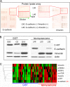
Fig. 1.
Fig. 1.
E-cadherin expression in leiomyosarcoma. A, reverse-phase protein lysate array analysis revealed the expression of E-cadherin in some leiomyosarcomas. Protein extract for each sample in six 2-fold serial dilutions was printed in triplicate. Results of E-cadherin and vimentin detection are shown with two examples (L46 and L50) highlighted. B, representative Western blotting validated the results of reverse-phase protein lysate array. In GIST samples G80, G2, and G19, vimentin and c-KIT are positive, and E-cadherin is negative (left panel). Leiomyosarcoma samples L46 and L92 show positive E-cadherin and negative vimentin, and L50 shows negative E-cadherin and positive vimentin (right panel). C, heat map from clustering analysis of the protein expression in leiomyosarcomas and GISTs by protein lysate array profiling. E-cadherin protein expression is significantly higher in a subset of leiomyosarcomas as indicated by the red color code. PDGFR, PDGF receptor; p-mTOR, phosphorylated mammalian target of rapamycin.
Fig. 2.
Fig. 2.
Validation of epithelial differentiation in leiomyosarcoma and analysis of clinical significance. A, epithelial differentiation in leiomyosarcoma detected by immunohistochemical staining on TMA. The upper panel includes examples of hematoxylin and eosin staining (left) and E-cadherin staining (right) in leiomyosarcoma. The lower panels show high magnification images of protein expression of E-cadherin, EMA, AE1/AE3, and pan-CK in leiomyosarcoma. B, leiomyosarcoma patients with positive E-cadherin protein expression had significantly better total survival than did those with no E-cadherin expression. C, leiomyosarcoma patients with higher mRNA expression of epithelial marker genes (E group) showed better survival than did those with higher mRNA expression of mesenchymal cell marker genes (M group). Cum, cumulative.
Fig. 3.
Fig. 3.
Inverse correlation of E-cadherin expression with Slug. A, correlation of E-cadherin expression with EMT- or MErT-related genes at the RNA level in leiomyosarcoma. Red indicates positive correlation; green indicates negative correlation. The arrow shows where the Slug mRNA has significant inverse correlation with E-cadherin mRNA expression. B, E-cadherin mRNA expression has significant inverse correlation with Slug (left) and N-cadherin (right). C, E-cadherin protein expression has significant inverse correlation with Slug detected by immunohistochemical staining on TMA. Upper panel, various levels of expression of E-cadherin protein; lower panel, inverse expression of Slug protein in the same samples in the adjacent section. VIM, vimentin; DES, desmin; CI, confidence interval.
Fig. 4.
Fig. 4.
Exogenous modulation of Slug-regulated E-cadherin expression, cell proliferation, invasion, and migration in SK-LMS-1 cells. A, Western blots of the endogenous expression of E-cadherin, Slug, AE1/AE3, N-cadherin, and vimentin in SK-LMS-1, TC71, SW480, and SW620 cells. B, Slug siRNA transfection decreases Slug, increases E-cadherin, and decreases N-cadherin and vimentin as shown on Western blotting. C, the decrease of Slug significantly decreases cell proliferation. D, the decrease of Slug significantly decreases cell invasion. E, the decrease of Slug significantly decreases cell migration. F, increased Slug decreases E-cadherin expression in SK-LMS-1 leiomyosarcoma cells. Western blot shows that Slug overexpression decreases E-cadherin and increases N-cadherin and vimentin. Cont, control.

References

    1. Alves C. C., Carneiro F., Hoefler H., Becker K. F. (2009) Role of the epithelial-mesenchymal transition regulator Slug in primary human cancers. Front. Biosci. 14, 3035–3050 - PubMed
    1. Olmeda D., Moreno-Bueno G., Flores J. M., Fabra A., Portillo F., Cano A. (2007) SNAI1 is required for tumor growth and lymph node metastasis of human breast carcinoma MDA-MB-231 cells. Cancer Res. 67, 11721–11731 - PubMed
    1. Yang M. H., Chen C. L., Chau G. Y., Chiou S. H., Su C. W., Chou T. Y., Peng W. L., Wu J. C. (2009) Comprehensive analysis of the independent effect of twist and snail in promoting metastasis of hepatocellular carcinoma. Hepatology 50, 1464–1474 - PubMed
    1. Saegusa M., Hashimura M., Kuwata T., Okayasu I. (2009) Requirement of the Akt/beta-catenin pathway for uterine carcinosarcoma genesis, modulating E-cadherin expression through the transactivation of slug. Am. J. Pathol. 174, 2107–2115 - PMC - PubMed
    1. Chen H., Zhu G., Li Y., Padia R. N., Dong Z., Pan Z. K., Liu K., Huang S. (2009) Extracellular signal-regulated kinase signaling pathway regulates breast cancer cell migration by maintaining slug expression. Cancer Res. 69, 9228–9235 - PMC - PubMed

Publication types

MeSH terms