

Isolation and perivascular localization of mesenchymal stem cells from mouse brain
- PMID: 20651630
- PMCID: PMC3644957
- DOI: 10.1227/01.NEU.0000377859.06219.78
Isolation and perivascular localization of mesenchymal stem cells from mouse brain
Abstract
Background: Although originally isolated from the bone marrow, mesenchymal stem cells (MSCs) have recently been detected in other tissues. However, little is known about MSCs in the brain.
Objective: To determine the extent to which cells with the features of MSCs exist in normal brain tissue and to determine the location of these cells in the brain.
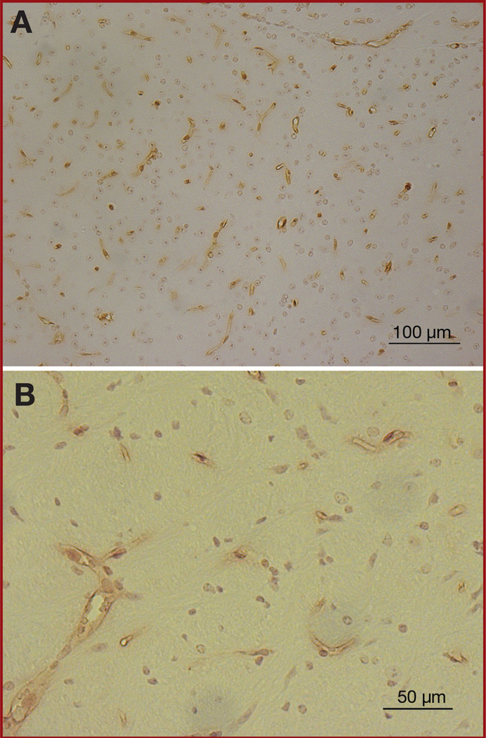
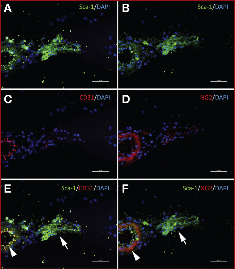
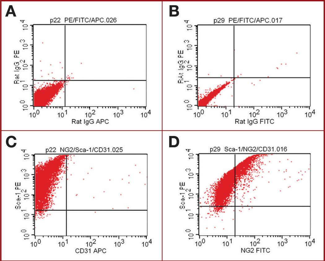
Methods: Single-cell suspensions from mouse brains were cultured according to the same methods used for culturing bone marrow-derived MSCs (BM-MSCs). These brain-derived cells were analyzed by fluorescence-activated cell sorting for surface markers associated with BM-MSCs (stem cell antigen 1 [Sca-1+], CD9+, CD45-, CD11b-, and CD31-). Brain-derived cells were exposed to mesenchymal differentiation conditions. To determine the locations of these cells within the brain, sections of normal brains were analyzed by immunostaining for Sca-1, CD31, and nerve/glial antigen 2.
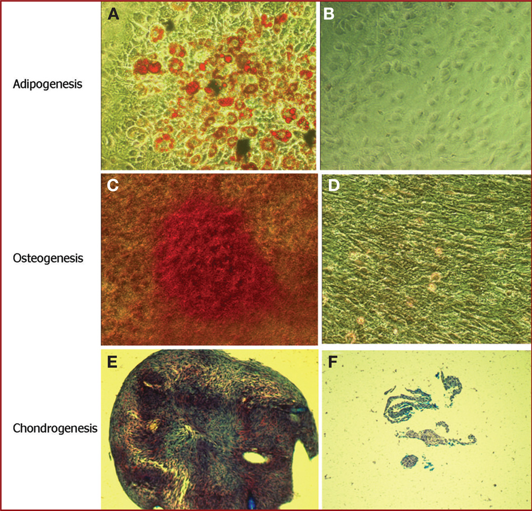
Results: Cells morphologically similar to mouse BM-MSCs were identified and called brain-derived MSCs (Br-MSCs). Fluorescence-activated cell sorting indicated that the isolated cells had a surface marker profile similar to BM-MSCs, ie, Sca-1V+, CD9+, CD45-, and CD11b-. Like BM-MSCs, Br-MSCs were capable of differentiation into adipocytes, osteocytes, and chondrocytes. Immunostaining indicated that Sca-1+ Br-MSCs are located around blood vessels and may represent progenitor cells that serve as a source of mesenchymal elements (eg, pericytes) within the brain.
Conclusion: Our results indicate that cells similar to BM-MSCs exist in the brain. These Br-MSCs appear to be located within the vascular niche and may provide the mesenchymal elements of this niche. Because MSCs may be part of the cellular response to tissue injury, Br-MSCs may represent targets in the therapy of pathological processes such as stroke, trauma, and tumorigenesis.
Figures


References
-
- Friedenstein AJ, Chailakhyan RK, Gerasimov UV. Bone marrow osteogenic stem cells: in vitro cultivation and transplantation in diffusion chambers. Cell Tissue Kinet. 1987;20(3):263–272. - PubMed
-
- Orkin SH. Hematopoietic stem cells: molecular diversification and developmental interrelationships. In: Marshak DR, Gardner RL, Gottlieb D, editors. Stem Cell Biology. Cold Spring Harbor, NY: Cold Spring Harbor Laboratory Press; 2001. pp. 289–306.
-
- Keller G. The hemangioblast. In: Marshak DR, Gardner RL, Gottlieb D, editors. Stem Cell Biology. Cold Spring Harbor, NY: Cold Spring Harbor Laboratory Press; 2001. pp. 329–348.
-
- Pittenger MF, Marshak DR. Mesenchymal stem cells of human adult bone marrow. In: Marshak DR, Gardner RL, Gottlieb D, editors. Stem Cell Biology. Vol. 16. Cold Spring Harbor, NY: Cold Spring Harbor Laboratory Press; 2001. pp. 349–373.
-
- Friedenstein AJ. Marrow stromal fibroblasts. Calcif Tissue Int. 1995;56:S17.
Publication types
MeSH terms
Grants and funding
- CA-16672/CA/NCI NIH HHS/United States
- P30 CA016672/CA/NCI NIH HHS/United States
- CA-1094551/CA/NCI NIH HHS/United States
- R01 CA109451/CA/NCI NIH HHS/United States
- P50 CA116199/CA/NCI NIH HHS/United States
- P50 CA 127001/CA/NCI NIH HHS/United States
- CA-49639/CA/NCI NIH HHS/United States
- R01 CA115729/CA/NCI NIH HHS/United States
- P01 CA049639/CA/NCI NIH HHS/United States
- P50 CA127001/CA/NCI NIH HHS/United States
- CA-116199/CA/NCI NIH HHS/United States
- CA-55164/CA/NCI NIH HHS/United States
- P01 CA055164/CA/NCI NIH HHS/United States
LinkOut - more resources
Full Text Sources
Research Materials
Miscellaneous
