

The REGENERATION of TOOTH ENAMEL
Dimens Dent Hyg.
2009 Aug.
No abstract available
Figures
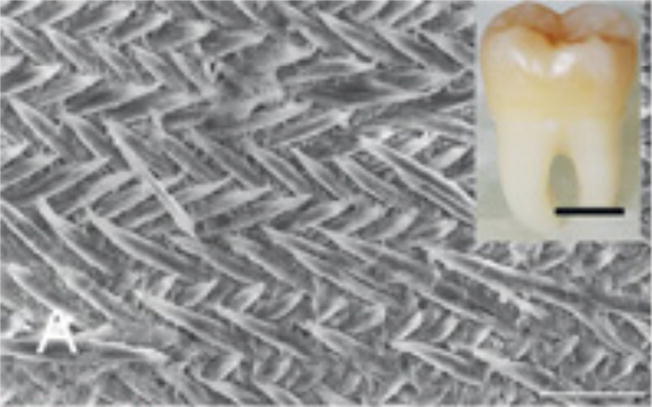
Figure 1A: Scanning electron microscope (SEM) images of etched surface of tooth enamel (mouse incisor) showing the interwoven arrangement of the prisms (scale 10 μm). Inset: A human third molar 6 (scale 1 cm). Figure 1B: Higher magnification SEM image showing the calcium fluoridated hydroxyapatite crystals arranged in parallel bundles within the prisms (scale 1 μm). Figure 1C: Transmission electron micrograph of a section of forming enamel (mouse molars) showing the arrangement black lines) within the protein-rich organic matrix (white circles) (scale 50 nanometers).
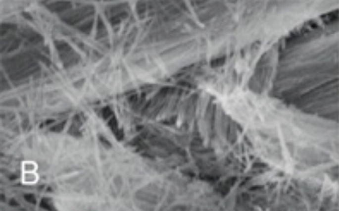
Figure 1A: Scanning electron microscope (SEM) images of etched surface of tooth enamel (mouse incisor) showing the interwoven arrangement of the prisms (scale 10 μm). Inset: A human third molar 6 (scale 1 cm). Figure 1B: Higher magnification SEM image showing the calcium fluoridated hydroxyapatite crystals arranged in parallel bundles within the prisms (scale 1 μm). Figure 1C: Transmission electron micrograph of a section of forming enamel (mouse molars) showing the arrangement black lines) within the protein-rich organic matrix (white circles) (scale 50 nanometers).
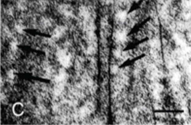
Figure 1A: Scanning electron microscope (SEM) images of etched surface of tooth enamel (mouse incisor) showing the interwoven arrangement of the prisms (scale 10 μm). Inset: A human third molar 6 (scale 1 cm). Figure 1B: Higher magnification SEM image showing the calcium fluoridated hydroxyapatite crystals arranged in parallel bundles within the prisms (scale 1 μm). Figure 1C: Transmission electron micrograph of a section of forming enamel (mouse molars) showing the arrangement black lines) within the protein-rich organic matrix (white circles) (scale 50 nanometers).
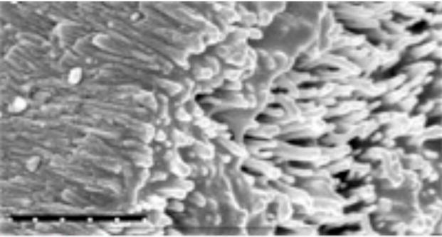
SEM of enamel-like hydroxy apatite crystals grown in the laboratory using the cation selective membrane system 12 (scale 1 μm).
References
-
- Nanci A. Ten Cate’s Oral Histology Development, Structure, and Function. St. Louis: Mosby Elsevier; 2008. Enamel: composition, formation, and structure; pp. 141–190.
-
- Thesleff I. Tooth morphogenesis. Adv Dent Res. 1995;9(3 Suppl):12. - PubMed
-
- Moradian-Oldak J, Paine ML. Mammalian. Enamel formation. In: Astrid S, Sigel H, Sigel RKO, editors. Metal Ions In Life Sciences. Chichester, United Kingdom: John Wiley & Sons Ltd; 2008. pp. 507–546.
-
- Simmer J, Hu J. Dental enamel formation and its impact on clinical dentistry. J Dent Educ. 2001;65:896–905. - PubMed
-
- Aoba T, Fejerskov O. Dental fluorosis: chemistry and biology. Crit Rev Oral Biol Med. 2002;13:155–170. - PubMed
Grants and funding
LinkOut - more resources
Full Text Sources