

Failed Switching off in the MIBI-Parathyroid Scintigraphy in a Dialyzed Patient with Secondary Hyperparathyroidism Responsive to Cinacalcet Therapy
- PMID: 20652073
- PMCID: PMC2905699
- DOI: 10.1155/2010/206801
Failed Switching off in the MIBI-Parathyroid Scintigraphy in a Dialyzed Patient with Secondary Hyperparathyroidism Responsive to Cinacalcet Therapy
Abstract
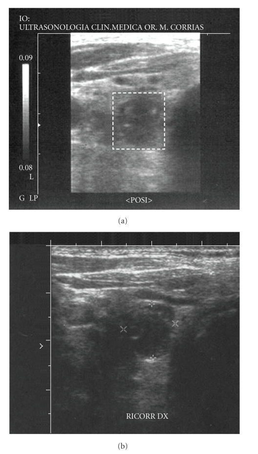
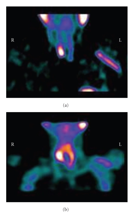
The aims of your case report is to show the predictivity of (99m)Tc-sestamibi (MIBI) scintigraphy and doppler ultrasound imaging on secondary hyperparathyroidism (SHPT) in a patient responsive to calcimimetic treatment. Moreover, it has been reported that calcimimetic has great potential in reducing the volume of the parathyroid gland. On the other hand, the MIBI scintigraphy is considered a crucial diagnostic procedure to monitor the response to therapy in terms of turnover and cellular metabolism; whereas, ultrasound to monitor the volume variation in response to treatment. It is described the case of a 73-year-old man on hemodialysis from 1995 for ESRD. Within 2 years the patient gradually developed SHPT with progressively increased iPTH up to 1,000 rhog/ml. The ultrasound, highlighted the presence of two parathyroid hyperplasia, confirmed by scintigraphy, showing focal increase uptake of sestamibi in the same anatomical areas. As a result of the patient's refusal to perform a parathyroidectomy, cinacalcet, was administered (65 mg overage daily dose). After a year of treatment, there was a striking decrease of iPTH (from 1300 to 57 rhog/ml, -95%); but, on the contrary to expectations, this positive metabolic outcome, was not followed by parathyroid changes in ultrasound and scintigraphic findings.
Similar articles
-
Preoperative evaluation of hyperparathyroidism: the role of dual-phase parathyroid scintigraphy and ultrasound imaging.Ann Nucl Med. 2008 Feb;22(2):123-31. doi: 10.1007/s12149-007-0086-z. Epub 2008 Mar 3. Ann Nucl Med. 2008. PMID: 18311537
-
Sestamibi scintigraphy, topography, and histopathology of parathyroid glands in secondary hyperparathyroidism.Am J Kidney Dis. 2006 Oct;48(4):638-44. doi: 10.1053/j.ajkd.2006.06.010. Am J Kidney Dis. 2006. PMID: 16997060 Clinical Trial.
-
Diagnostic performance of ultrasonography, dual-phase 99mTc-MIBI scintigraphy, early and delayed 99mTc-MIBI SPECT/CT in preoperative parathyroid gland localization in secondary hyperparathyroidism.BMC Med Imaging. 2020 Aug 3;20(1):91. doi: 10.1186/s12880-020-00490-3. BMC Med Imaging. 2020. PMID: 32746794 Free PMC article.
-
Predicting the effect of intravenous calcitriol on parathyroid gland activity using double-phase technetium Tc 99m-sestamibi scintigraphy.Am J Kidney Dis. 2004 Sep;44(3):476-80. Am J Kidney Dis. 2004. PMID: 15332220
-
[Usefulness of imaging techniques in secondary hyperparathyroidism].Nefrologia. 2010;30(2):158-67. doi: 10.3265/Nefrologia.pre2010.Jan.10231. Epub 2010 Jan 21. Nefrologia. 2010. PMID: 20098464 Review. Spanish.
References
-
- Sprague SM, Coyne D. Control of secondary hyperparathyroidism by vitamin D receptor agonists in chronic kidney disease. Clinical Journal of the American Society of Nephrology. 2010;5(3):512–518. - PubMed
-
- Bover J, Aguilar A, Baas JP, et al. Calcimimetics in the chronic kidney disease-mineral and bone disorder. International Journal of Artificial Organs. 2009;32(2):108–121. - PubMed
-
- Fuster D, Ybarra J, Torregrosa JV, et al. Double-phase parathyroid 99mTc-sestamibi scintigraphy in chronic haemodialysis patients: correlation with biochemical markers of parathyroid function. Nuclear Medicine Communications. 2003;24(1):85–90. - PubMed
-
- Gotway MB, Reddy GP, Webb WR, Morita ET, Clark OH, Higgins CB. Comparison between MR imaging and 99mTc MIBI scintigraphy in the evaluation of recurrent or persistent hyperparathyroidism. Radiology. 2001;218(3):783–790. - PubMed
-
- Olaizola I, Zingraff J, Heuguerot C, et al. [(99m)Tc]-sestamibi parathyroid scintigraphy in chronic haemodialysis patients: static and dynamic explorations. Nephrology Dialysis Transplantation. 2000;15(8):1201–1206. - PubMed
Publication types
LinkOut - more resources
Full Text Sources
Research Materials
