. 2009 Sep 16;4(12):1493-501.
doi: 10.1007/s11671-009-9426-3.

Nanoscale Visualization of Elastic Inhomogeneities at TiN Coatings Using Ultrasonic Force Microscopy

Affiliations

Nanoscale Visualization of Elastic Inhomogeneities at TiN Coatings Using Ultrasonic Force Microscopy

Ja Hidalgo et al. Nanoscale Res Lett. .

Abstract

Ultrasonic force microscopy has been applied to the characterization of titanium nitride coatings deposited by physical vapor deposition dc magnetron sputtering on stainless steel substrates. The titanium nitride layers exhibit a rich variety of elastic contrast in the ultrasonic force microscopy images. Nanoscale inhomogeneities in stiffness on the titanium nitride films have been attributed to softer substoichiometric titanium nitride species and/or trapped subsurface gas. The results show that increasing the sputtering power at the Ti cathode increases the elastic homogeneity of the titanium nitride layers on the nanometer scale. Ultrasonic force microscopy elastic mapping on titanium nitride layers demonstrates the capability of the technique to provide information of high value for the engineering of improved coatings.

Keywords: Nanomechanics; PVD nanostructured coatings; Scanning probe microscopy; TiN; Ultrasonic force microscopy.


Figures
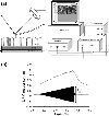
Figure 1
Figure 1
aSet-up for the UFM measurements;bTypical UFM cantilever response when a modulated ultrasonic excitation of 4 MHz with maximum amplitude Am = 8 Vppis applied to the piezo beneath the TiN sample (S-UFM mode), being the initial tip-sample set-point force (in the absence of ultrasound) of ≈70 nN
Figure 2
Figure 2
aXRD patterns (θ-2θBragg–Brentano scan) of TiN deposited on SS304 with different sputtering power (WS) andbschematic representation of the δ-TiN/α-Ti/SS304 system with the TiN grains growing in a specific direction
Figure 3
Figure 3
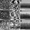
SEM (a–a″), AFM (b–b″) topographic images and SEM cross sectional view (c–c″) of the TiN film on SS304 produced withWS = 100 W (a–c), 150 W (a′–c′) and 200 W (a″–c″). Thegrey-scale range in AFM images (b–b″) is 112, 160 and 125 nm, respectively
Figure 4
Figure 4
TiN film obtained atWS = 100 W:aTopography in AFM contact mode. Surface area: (5 × 5) 03BCm2;Grey-scale range: 466 nm.bUFM image simultaneously recorded with (a).cTopography in AFM contact mode recorded over the square region in (a). Surface area: (1 × 1) μm2Grey-scale range: 72 nm.dUFM image simultaneously recorded with (c), over the region squared in (b).eTopographic andfelastic profile along thelinesindicated in (c) and (d), respectively
Figure 5
Figure 5
aAFM topographic image. Surface area: (500 × 500) nm2Grey-scale range: 58 nm.bDerivative image of (a).cUFM image simultaneously recorded with (a), over the region near to thedarker grain(iii) in 4d
Figure 6
Figure 6
TiN film obtained atWS = 150 W:aTopography in AFM contact mode. Surface area: (5 × 5) μm2Grey-scale range: 288 nm.bUFM image simultaneously recorded with (a).cTopography in AFM contact mode. Surface area: (1 × 1) μm2.Grey-scale range: 64 nm.dDerivative image of (c).eUFM image over the squared region in (a,b)
Figure 7
Figure 7
TiN film obtained atWS = 200 W.aTopography in AFM contact mode. Surface area: (5 × 5) μm2Grey-scale range: 119 nm.bUFM image simultaneously recorded with (a). Thearrowsindicate softer UFM regions and their corresponding location in the AFM image

References

    1. Steyer PH, Mege A, Pech D, Mendibide C, Fontaine J, Pierson J-F, Esnouf C, Goudeau P. Surf. 2008. p. 2268. COI number [1:CAS:528:DC%2BD1cXit1SqsL8%3D] - DOI
    1. V. Karagkiozaki, S. Logothetidis, N. Kalfagiannis, S. Lousinian, G. Giannoglou, Nanomedicine. doi:10.1016/j.nano.2008.07.005 (2008) - PMC - PubMed
    1. Kiran MSRN, Krishna MG, Padmanabhan KA. Appl. Surf. Sci. 2008. p. 1934. COI number [1:CAS:528:DC%2BD1cXhsVChs7nM]; Bibcode number [2008ApSS..255.1934K] - DOI
    1. M. Stueber, H. Holleck, H. Leiste, K. Seemann, S. Ulrich, C. Ziebert, J. Alloys Compd. doi:10.1016/j.jallcom.2008.08.133 (2008)
    1. Krella A, Czyzniewski A. Wear. 2008. p. 963. COI number [1:CAS:528:DC%2BD1cXmvFOmtr0%3D] - DOI

LinkOut - more resources