Review
. 2010 Sep;32(3):305-17.
doi: 10.1007/s00281-010-0217-9. Epub 2010 Jul 22.

Intravital imaging of anti-tumor immune response and the tumor microenvironment

Affiliations
Review

Intravital imaging of anti-tumor immune response and the tumor microenvironment

Tomasz Zal et al. Semin Immunopathol. 2010 Sep.

Abstract

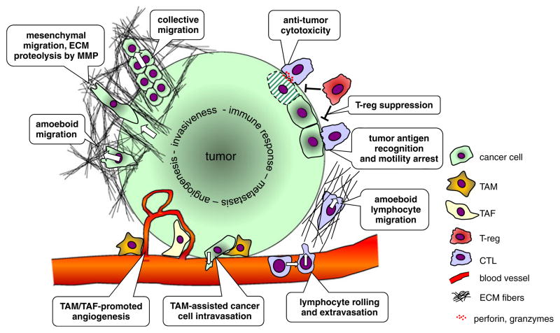
Tumor growth, invasiveness, and metastasis are dynamic processes involving cancer interactions with the extracellular matrix, the vasculature, and various types of non-cancerous host cells that form the tumor stroma. An often-present stromal component is the immune cells, such as tumor-associated myeloid and lymphocytic infiltrates, yet endogenous anti-tumor immune responses are typically ineffective in tumor rejection and may even contribute to the progression of some cancers. How exactly cancer cells interact with the stroma and invade healthy tissues while avoiding anti-tumor immune responses, and which interactions should be targeted for anti-tumor therapy, can now be studied by minimally invasive observation using multiphoton and other low impact confocal microscopy techniques and fluorescent animal tumor models. Intravital video microscopy has already been instrumental in defining the roles and modes of cellular motility in the angiogenic process and during tissue invasion at the tumor margin. In the hands of cancer immunologists, intravital video microscopy is beginning to unravel the complexity of effector and suppressory lymphocytic interactions in tumors and in the draining lymphoid organs. As the intravital microscopy approach is beginning to move beyond fundamental description and into analyzing the molecular underpinnings of cell's dynamics, future technical advances will undoubtedly provide yet deeper insight while stitching together a systems dynamics view of cancer-host interactions that will keep on inspiring cancer researchers and therapists.


Figures

Fig. 1
Fig. 1
Schematic representation of cell–cell and cell–matrix interactions in the tumor microenvironment. At the invasive edge, cancer cells move through the extracellular matrix (ECM) outwards into the surrounding tissue. Two types of translational motility can be distinguished based on cell morphological changes and its interactions with the fibrillar collagen networks. Mesenchymal motility is typical for connective tissue cells and is characterized by matrix metalloproteinase—mediated ECM degradation at the leading cell edge, which leaves tubular tracks that can guide other cells. Collective migration can be regarded as cooperative mesenchymal motility. Amoeboid motility is typical for lymphocytes and occurs without ECM degradation by transient formation of lamellipodia, focal cell attachment, and uropod retraction. Under certain conditions, cancer cell motility can undergo a mesenchymal-amoeboid transition. Tumor-associated macrophages (TAM) and tumor-associated fibroblasts contribute to tumor angiogenesis, and TAM can assist cancer cells intravasating into the lumen of blood vessels. Lymphocyte interactions with tumors involve recruitment from the circulation via integrin-mediated rolling followed by extravasation. Upon amoeboid migration into the tumor mass, tumor antigen-specific cytotoxic T lymphocytes (CTL) can form stable contacts with cancer cells and kill the cells by apoptosis. Regulatory FoxP3+ CD4+ T cells can interfere with CTL effector function by inhibiting cytotoxic granule release
Fig. 2
Fig. 2
Live imaging of T cell recruitment to a lung sarcoma tumor. A dual reporter hCD2p-DsRed × CD8β-YFP mouse was implanted s.c. with the methylcholanthrene (MCA) sarcoma tumor on day 0 to mimic a primary tumor. On day 7, MCA-mCerulean cells were injected i.v. to induce lung metastasis. Intravital microscopy of the lungs was performed on day 8 to visualize cytotoxic T lymphocytes (CTL) recruitment to micrometastases. Blue: MCA-mCerulean tumor, solid arrow; red: DsRed is expressed in all T cells [81]; green: CD8β-YFP chains are expressed on the cell surface only in CD8+ T cells by pairing with CD8α chains [82]. Cell tracking analysis was performed on selected CD8+ T cells. Note the two CD8+ T cells (open arrows) that extravasate next to the tumor cell at 6 min 20 s and migrate as indicated by the white tracks. Data acquisition: Leica SP5RS resonant scanning confocal microscope, ×20 NA 0.95 water immersion objective, 458, 514, and 543 nm excitation. The images represent maximum intensity projections from image stack spaced at z=3 μm. Temporal resolution, 20 s. The data provides an example of the benefits of fast resonant scanning confocal microscopy and fluorescent reporter mice for intravital imaging of CTL activity in experimental lung metastases

Similar articles

Cited by

References

    1. Peggs KS, Quezada SA, Korman AJ, Allison JP. Principles and use of anti-CTLA4 antibody in human cancer immunotherapy. Curr Opin Immunol. 2006;18:206–213. - PubMed
    1. Sakaguchi S, Sakaguchi N, Asano M, Itoh M, Toda M. Immunologic self-tolerance maintained by activated T cells expressing IL-2 receptor alpha-chains (CD25). Breakdown of a single mechanism of self-tolerance causes various autoimmune diseases. J Immunol. 1995;155:1151–1164. - PubMed
    1. Shimizu J, Yamazaki S, Sakaguchi S. Induction of tumor immunity by removing CD25+CD4+ T cells: a common basis between tumor immunity and autoimmunity. J Immunol. 1999;163:5211–5218. - PubMed
    1. Fontenot JD, Gavin MA, Rudensky AY. Foxp3 programs the development and function of CD4+CD25+ regulatory T cells. Nat Immunol. 2003;4:330–336. - PubMed
    1. Xu C, Zipfel W, Shear JB, Williams RM, Webb WW. Multiphoton fluorescence excitation: new spectral windows for biological nonlinear microscopy. Science. 1996;93:10763–10768. - PMC - PubMed

Publication types

MeSH terms

Substances