

A role of diffusion tensor imaging in movement disorder surgery
- PMID: 20652606
- PMCID: PMC2991222
- DOI: 10.1007/s00701-010-0742-2
A role of diffusion tensor imaging in movement disorder surgery
Abstract
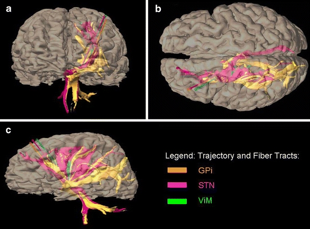
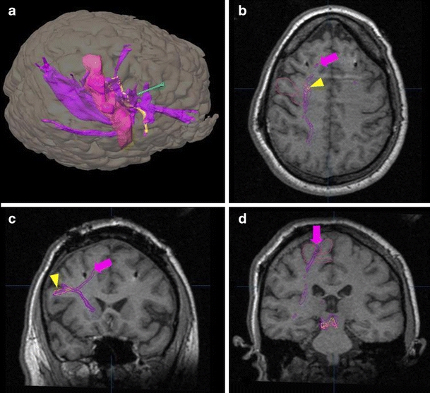
The safe and reversible nature of deep brain stimulation (DBS) has allowed movement disorder neurosurgery to become commonplace throughout the world. Fundamental understanding of individual patient's anatomy is critical for optimizing the effects and side effects of DBS surgery. Three patients undergoing stereotactic surgery for movement disorders, at the institution's intraoperative magnetic resonance imaging operating suite, were studied with fiber tractography. Stereotactic targets and fiber tractography were determined on preoperative magnetic resonance imagings using the Schaltenbrand-Wahren atlas for definition in the BrainLab iPlan software (BrainLAB Inc., Feldkirchen, Germany). Subthalamic nucleus, globus pallidus interna, and ventral intermediate nucleus targets were studied. Diffusion tensor imaging parameters used ranged from 2 to 8 mm for volume of interest in the x/y/z planes, fiber length was kept constant at 30 mm, and fractional anisotropy threshold varied from 0.20 to 0.45. Diffusion tensor imaging tractography allowed reliable and reproducible visualization and correlation between frontal eye field, premotor, primary motor, and primary sensory cortices via corticospinal tracts and corticopontocerebellar tracts. There is an apparent increase in the number of cortical regions targeted by the fiber tracts as the region of interest is enlarged. This represents a possible mechanism of the increased effects and side effects observed with higher stimulation voltages. Currently available diffusion tensor imaging techniques allow potential methods to characterize the effects and side effects of DBS. This technology has the potential of being a powerful tool to optimize DBS neurosurgery.
Figures


Similar articles
-
Diffusion tensor imaging (DTI) and colored fractional anisotropy (FA) mapping of the subthalamic nucleus (STN) and the globus pallidus interna (GPi).Acta Neurochir (Wien). 2010 Dec;152(12):2079-84. doi: 10.1007/s00701-010-0813-4. Epub 2010 Oct 3. Acta Neurochir (Wien). 2010. PMID: 20890778 Free PMC article.
-
Tractography-assisted deep brain stimulation of the superolateral branch of the medial forebrain bundle (slMFB DBS) in major depression.Neuroimage Clin. 2018 Aug 14;20:580-593. doi: 10.1016/j.nicl.2018.08.020. eCollection 2018. Neuroimage Clin. 2018. PMID: 30186762 Free PMC article. Clinical Trial.
-
Correlation of diffusion tensor tractography and intraoperative macrostimulation during deep brain stimulation for Parkinson disease.J Neurosurg. 2014 Oct;121(4):929-35. doi: 10.3171/2014.6.JNS131673. Epub 2014 Jul 25. J Neurosurg. 2014. PMID: 25061862
-
Integrating diffusion tensor imaging-based tractography into deep brain stimulation surgery: a review of the literature.Stereotact Funct Neurosurg. 2014;92(5):282-90. doi: 10.1159/000362937. Epub 2014 Sep 18. Stereotact Funct Neurosurg. 2014. PMID: 25248076 Review.
-
Diffusion Tensor Imaging of the Basal Ganglia for Functional Neurosurgery Applications.Prog Neurol Surg. 2018;33:62-79. doi: 10.1159/000480766. Epub 2018 Jan 12. Prog Neurol Surg. 2018. PMID: 29332074 Review.
Cited by
-
Targeting of the dentato-rubro-thalamic tract for MR-guided focused ultrasound treatment of essential tremor.Neuroradiol J. 2019 Dec;32(6):401-407. doi: 10.1177/1971400919870180. Epub 2019 Aug 13. Neuroradiol J. 2019. PMID: 31407957 Free PMC article.
-
Diffusion MRI tractography for improved transcranial MRI-guided focused ultrasound thalamotomy targeting for essential tremor.Neuroimage Clin. 2018 May 9;19:572-580. doi: 10.1016/j.nicl.2018.05.010. eCollection 2018. Neuroimage Clin. 2018. PMID: 29984165 Free PMC article.
-
Estimating Brain Connectivity With Diffusion-Weighted Magnetic Resonance Imaging: Promise and Peril.Biol Psychiatry Cogn Neurosci Neuroimaging. 2020 Sep;5(9):846-854. doi: 10.1016/j.bpsc.2020.04.009. Epub 2020 Apr 27. Biol Psychiatry Cogn Neurosci Neuroimaging. 2020. PMID: 32513555 Free PMC article. Review.
-
In vivo Exploration of the Connectivity between the Subthalamic Nucleus and the Globus Pallidus in the Human Brain Using Multi-Fiber Tractography.Front Neuroanat. 2017 Jan 19;10:119. doi: 10.3389/fnana.2016.00119. eCollection 2016. Front Neuroanat. 2017. PMID: 28154527 Free PMC article.
-
White matter tractography for neurosurgical planning: A topography-based review of the current state of the art.Neuroimage Clin. 2017 Jun 15;15:659-672. doi: 10.1016/j.nicl.2017.06.011. eCollection 2017. Neuroimage Clin. 2017. PMID: 28664037 Free PMC article. Review.
