Review
. 2011 Jul;41(2):257-69.
doi: 10.1007/s00726-010-0676-2. Epub 2010 Jul 21.

ADP-ribosylation of arginine

Affiliations
Review

ADP-ribosylation of arginine

Sabrina Laing et al. Amino Acids. 2011 Jul.

Abstract

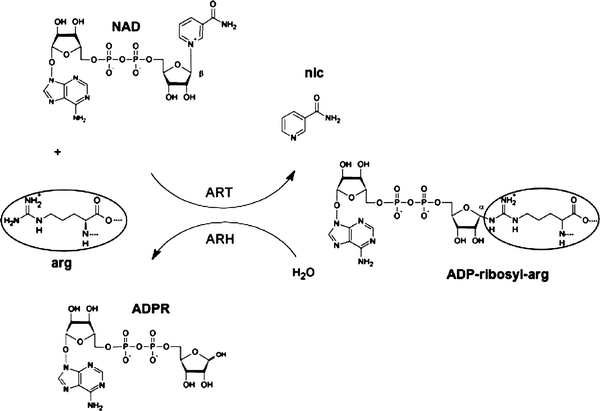
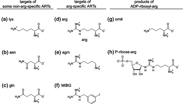
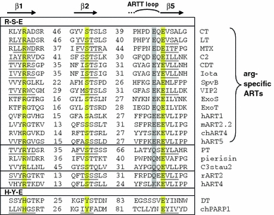
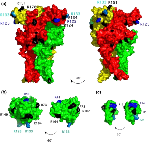
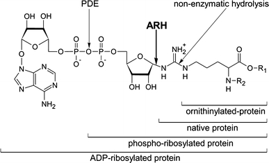
Arginine adenosine-5'-diphosphoribosylation (ADP-ribosylation) is an enzyme-catalyzed, potentially reversible posttranslational modification, in which the ADP-ribose moiety is transferred from NAD(+) to the guanidino moiety of arginine. At 540 Da, ADP-ribose has the size of approximately five amino acid residues. In contrast to arginine, which, at neutral pH, is positively charged, ADP-ribose carries two negatively charged phosphate moieties. Arginine ADP-ribosylation, thus, causes a notable change in size and chemical property at the ADP-ribosylation site of the target protein. Often, this causes steric interference of the interaction of the target protein with binding partners, e.g. toxin-catalyzed ADP-ribosylation of actin at R177 sterically blocks actin polymerization. In case of the nucleotide-gated P2X7 ion channel, ADP-ribosylation at R125 in the vicinity of the ligand-binding site causes channel gating. Arginine-specific ADP-ribosyltransferases (ARTs) carry a characteristic R-S-EXE motif that distinguishes these enzymes from structurally related enzymes which catalyze ADP-ribosylation of other amino acid side chains, DNA, or small molecules. Arginine-specific ADP-ribosylation can be inhibited by small molecule arginine analogues such as agmatine or meta-iodobenzylguanidine (MIBG), which themselves can serve as targets for arginine-specific ARTs. ADP-ribosylarginine specific hydrolases (ARHs) can restore target protein function by hydrolytic removal of the entire ADP-ribose moiety. In some cases, ADP-ribosylarginine is processed into secondary posttranslational modifications, e.g. phosphoribosylarginine or ornithine. This review summarizes current knowledge on arginine-specific ADP-ribosylation, focussing on the methods available for its detection, its biological consequences, and the enzymes responsible for this modification and its reversal, and discusses future perspectives for research in this field.


Figures

Fig. 1
Fig. 1
Schematic diagram of the enzyme catalyzed, reversible posttranslational modification of arginine by ADP-ribose. In the active centre of an ADP-ribosyltransferase (ART), NAD+ is brought into an extended conformation that permits the attack of the target arginine on the β-N-glycosidic bond between nicotinamide and the C1′-atom of the ribose group. This leads to the formation of ADP-ribosylarginine with C1′ in α-conformation, while nicotinamide is released. The native arginine can be recovered by the reverse reaction, catalyzed by an ADP-ribosylarginine hydrolase (ARH). This enzyme hydrolyses the α-glycosidic bond, releasing ADP-ribose
Fig. 2
Fig. 2
Monitoring ADP-ribosylation by specific and promiscuous ARTs by SDS-PAGE autoradiography. HEK-cell lysates were incubated for 10 min at 37°C with arginine-specific ARTs (ART2.2, SpvB, C2) or non-arginine-specific ARTs (C3stau2, C3bot, PT) in the presence of [32P]-NAD+. Proteins were size-fractionated by SDS-PAGE and visualized by Coomassie staining (a). SDS-resistant, i.e. covalently incorporated, radioactivity was detected by autoradiography (b). While most bacterial ARTs modify a single prominent band, mouse ART2.2 (lane 1) modifies many target proteins. Enzymes used for ADP-ribosylation: Mouse ART2.2 (ART2); Salmonella enterica SpvB toxin (SpvB); Clostridium botulinum C2 toxin (C2); Staphylococcus aureus exoenzyme C3stau2 (C3stau); Clostridium botulinum C3 toxin (C3bot); Pertussis toxin (PT)
Fig. 3
Fig. 3
Schematic diagrams of molecules relevant to ADP-ribosylation. ac Target amino acids with a terminal nitrogen group that can be modified by non-arginine specific ARTs; df guanidino group-containing targets of arginine-specific ARTs; g, h products of enzymatic and non-enzymatic hydrolysis of ADP-ribosylated-arginine. a lysine; b asparagine; c glutamine; d arginine; e agmatine; f MIBG (meta-iodobenzylguanidine); g ornithine; h phospho-ribosylarginine
Fig. 4
Fig. 4
Sequence alignment of arginine- and non-arginine-specific ARTs. The regions surrounding the three catalytically important residues on the β1, β2, and β5 strands are aligned (Hottiger et al. ; Koch-Nolte et al. ; Otto et al. 2005). The R-S-E and H-Y-E motifs characterizing the major ART families are highlighted in yellow. For the R-S-E ARTs, the residue in position −2 relative to the catalytic glutamic acid in β5 is highlighted in grey. Specificity for arginine is determined by the presence of glutamic acid at this position (R-S-EXE motif). Enzymes shown in the alignment: cholera toxin (CT); Escherichia coli heat labile enterotoxin (LT); Bacillus sphaericus mosquitocidal toxin (MTX); Clostridium botulinum C2 toxin (C2); Clostridium difcile ADP-ribosylating toxin (CDT); Clostridium perfringens iota toxin (Iota); Salmonella enterica SpvB toxin (SpvB); Bacillus cereus VIP2 toxin (VIP2); Pseudomonas aeruginosa exoenzyme S (ExoS); Pseudomonas aeruginosa exoenzyme T (ExoT); human ART1 (hART1); mouse ART2.2 (mART2.2); chicken ART4 (chART4); human ART5 (hART5); pertussis toxin (PT); Pieris rapae pierisin-1 (pierisin); Staphylococcus aureus exoenzyme C3stau2 (C3stau2); rat ART2 (rART2); human ART4 (hART4); diphtheria toxin (DT); chicken poly(ADP-ribose)transferase-1 (chPARP-1). Underlined sequences indicate residues confirmed to be in β-strands in the respective crystal-structures. The ARTT (ADP-ribosyltransferase turn-turn) loop is involved in target protein recognition and contributes to catalysis (Koch-Nolte et al. ; Menetrey et al. 2008)
Fig. 5
Fig. 5
Three-dimensional models of P2X7 (a), ras (b), and HNP-1 (c), highlighting arginine residues that are targets for ADP-ribosylation. Preferred (primary) ADP-ribosylation sites are highlighted in blue, secondary target residues in cyan. Other arginine residues that are not targets for ADP-ribosylation are shown in black. Two views of each molecule, rotated as indicated by the arrow, are shown. The representations were made using the PyMol Molecular Viewer software and are based on the following structures in the PDB database: 2KHT (HNP-1) and 121P (ras). For P2X7, arginine residues were projected onto the recently solved structure of zebrafish P2X4 (PDB: 3I5D)
Fig. 6
Fig. 6
Enzymatic and non-enzymatic processing of ADP-ribosylarginine. See text for details. ARH ADP-ribosylhydrolase, PDE phosphodiesterase

Similar articles

Cited by

References

    1. Adriouch S, Bannas P, Schwarz N, Fliegert R, Guse AH, Seman M, Haag F, Koch-Nolte F. ADP-ribosylation at R125 gates the P2X7 ion channel by presenting a covalent ligand to its nucleotide binding site. FASEB J. 2008;22:861–869. doi: 10.1096/fj.07-9294com. - DOI - PubMed
    1. Aktories K, Barth H. Clostridium botulinum C2 toxin—new insights into the cellular up-take of the actin-ADP-ribosylating toxin. Int J Med Microbiol. 2004;293:557–564. doi: 10.1078/1438-4221-00305. - DOI - PubMed
    1. Aktories K, Barmann M, Ohishi I, Tsuyama S, Jakobs KH, Habermann E. Botulinum C2 toxin ADP-ribosylates actin. Nature. 1986;322:390–392. doi: 10.1038/322390a0. - DOI - PubMed
    1. Altmeyer M, Messner S, Hassa PO, Fey M, Hottiger MO. Molecular mechanism of poly(ADP-ribosyl)ation by PARP1 and identification of lysine residues as ADP-ribose acceptor sites. Nucleic Acids Res. 2009;37:3723–3738. doi: 10.1093/nar/gkp229. - DOI - PMC - PubMed
    1. Bannas P, Adriouch S, Kahl S, Braasch F, Haag F, Koch-Nolte F. Activity and specificity of toxin-related mouse T cell ecto-ADP-ribosyltransferase ART2.2 depends on its association with lipid rafts. Blood. 2005;105:3663–3670. doi: 10.1182/blood-2004-08-3325. - DOI - PubMed

Publication types