. 2010 Sep;28(9):1661-73.
doi: 10.1002/stem.488.

FRS2α regulates Erk levels to control a self-renewal target Hes1 and proliferation of FGF-responsive neural stem/progenitor cells

Affiliations
Free PMC article

FRS2α regulates Erk levels to control a self-renewal target Hes1 and proliferation of FGF-responsive neural stem/progenitor cells

Takuya Sato et al. Stem Cells. 2010 Sep.
Free PMC article

Abstract

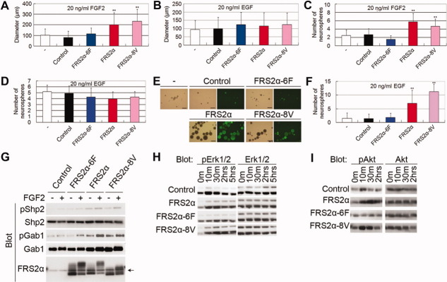
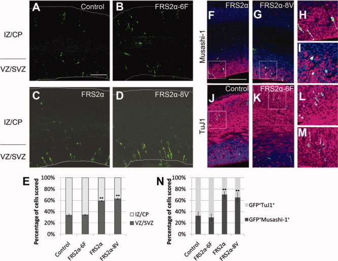
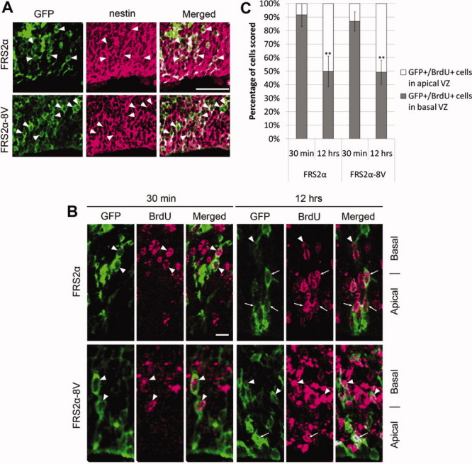
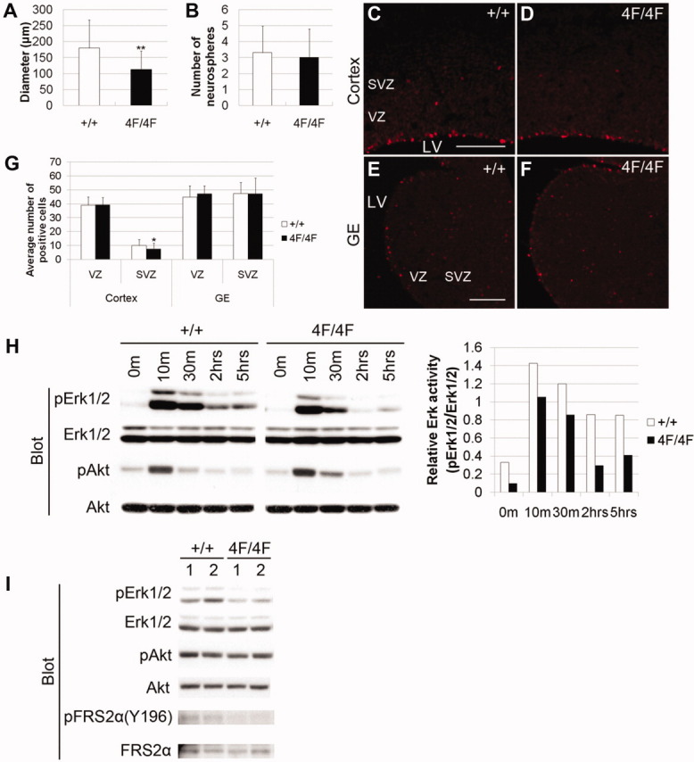
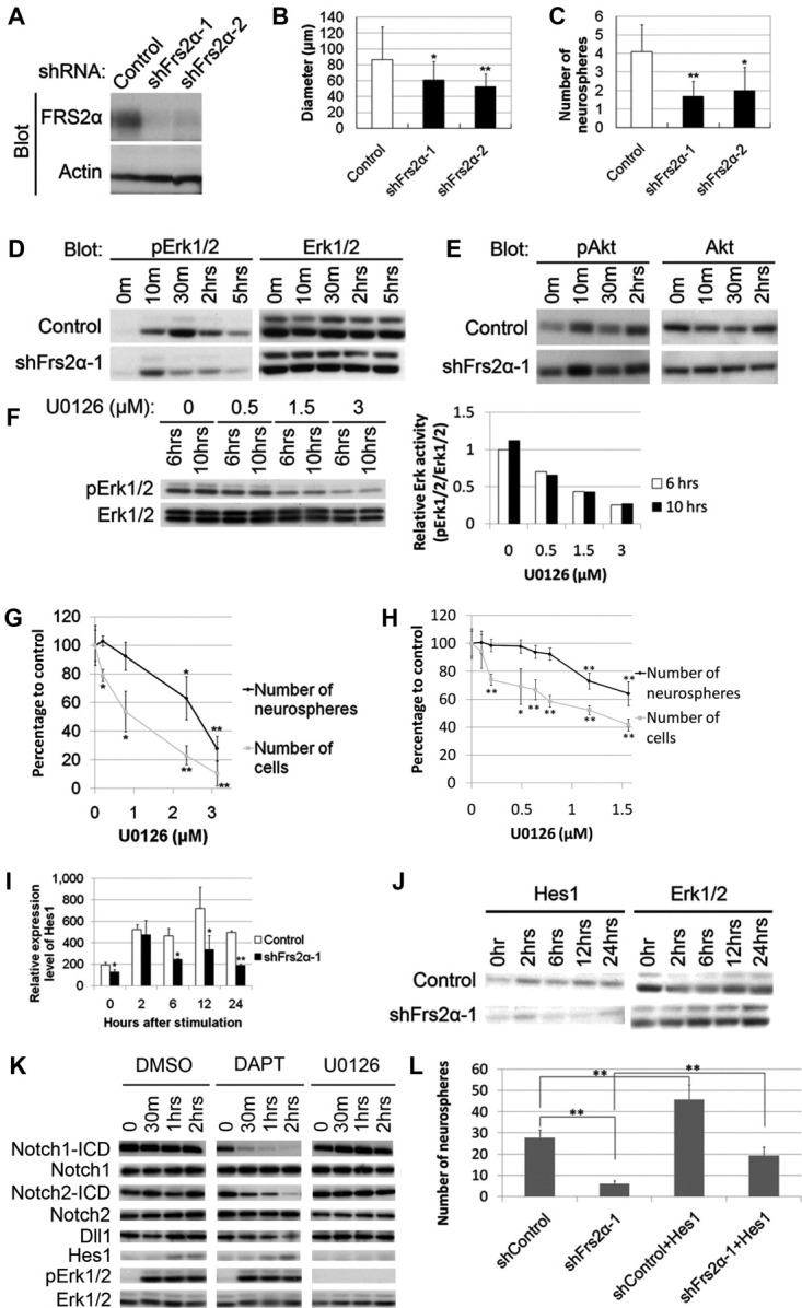
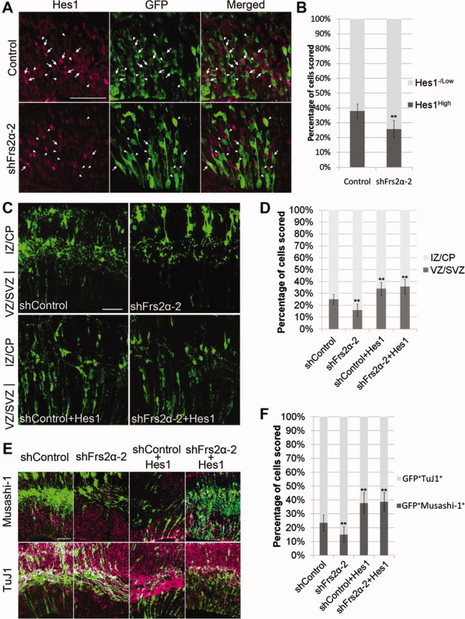
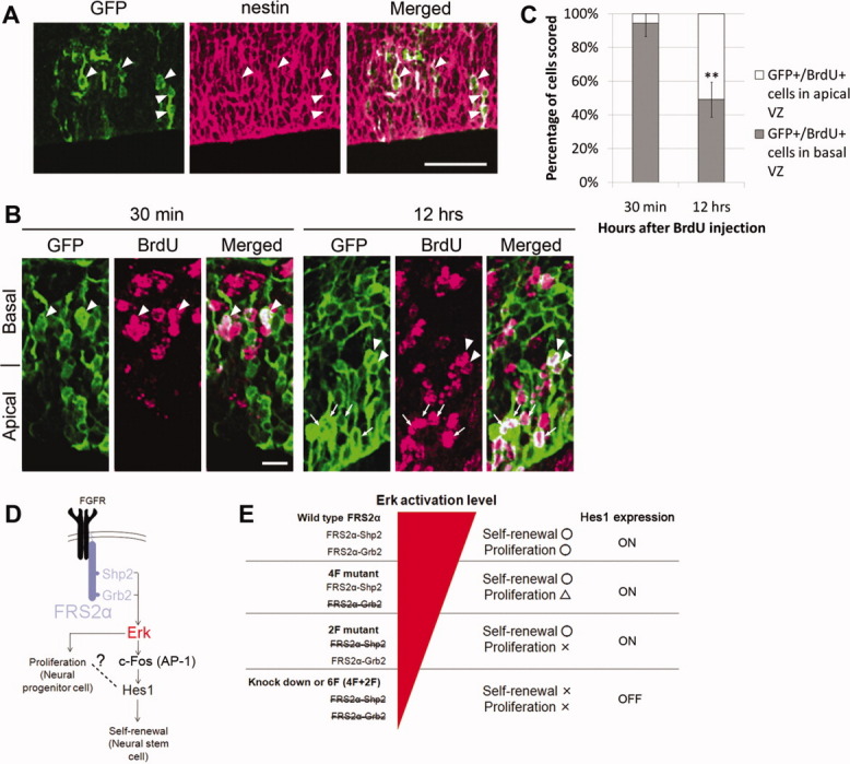
Fibroblast growth factor (FGF) is among the most common growth factors used in cultures to maintain self-renewal and proliferative capabilities of a variety of stem cells, including neural stem cells (NSCs). However, the molecular mechanisms underlying the control by FGF have remained elusive. Studies on mutant mice of FGF receptor substrate 2α (FRS2α), a central mediator for FGF signaling, combined with FRS2α knockdown or gain-of-function experiments, allowed us to dissect the role of FGF signaling for the self-renewal and proliferation of NSCs and to provide novel molecular mechanisms for them. We identified Hes1 as a novel self-renewal target of FGF-signaling. Quantitatively different levels of Erk activation mediated by FRS2α may regulate self-renewal of NSCs and proliferation of neural stem/progenitor cells (NSPCs); low levels of Erk activation are sufficient for the former, however, higher levels are required for maximum activity of the latter. Thus, FRS2α fine-tunes the FGF-signaling to control qualitatively different biological activities, self-renewal at least partly through Hes1 versus proliferation of NSPCs.


Figures

Figure 1
Figure 1
Overexpression of FRS2α promotes FGF2-induced proliferation and self-renewal of neural stem/progenitor cells (NSPCs) in vitro. (A–E): NSPCs were infected with retrovirus and cultured in the presence of FGF2 (A, C) or EGF (B, D). Then, the diameter (A, B) or number (C, D) of resulting neurospheres was determined. A minus (−) sign indicates that the virus was not infected. Scale bars = 500 μm (E). (F): EGF-induced tertiary neurospheres were produced from FGF2-induced secondary neurospheres, and the number of neurospheres was determined. Experiments were performed at least two times with similar results. (G–I): Neurospheres were starved and then stimulated with FGF2. Then, the lysates were subjected to immunoblotting. The arrow in (G) indicates nonspecific bands. Abbreviations: EGF, epidermal growth factor; FGF, fibroblast growth factor; FRS2α, FGF receptor substrate 2α.
Figure 2
Figure 2
Overexpression of FRS2α represses the differentiation of neural stem/progenitor cells (NSPCs) in the developing cortex. (A–D): Retroviruses were injected in utero into the telencephalic ventricles at E12.5. The embryos were then sacrificed at E15.5, and cortical sections were immunostained against GFP (green). (E): Quantification of the data presented in (A–D). In each condition, at least 183 cells were counted in 10 randomly selected areas. (F–M): Cortical sections of embryo infected with viruses were double-immunostained against GFP (green) and Musashi-1 (magenta) (F–I), or TuJ1 (magenta) (J–M). Nuclear staining is shown in blue. The panels (H), (I), (L), and (M) are higher magnifications of the regions indicated by rectangles in (F), (G), (J), and (K), respectively. (N): Quantification of the data presented in (F–M). In each condition, at least 64 cells were counted in 10 randomly selected areas. Scale bars = 50 μm. Abbreviations: CP, cortical plate; GFP, Green Fluorescent Protein; FRS2α, FGF receptor substrate 2α; IZ, intermediate zone; SVZ, subventricular zone; VZ, ventricular zone.
Figure 3
Figure 3
Cells expressing FRS2α or FRS2α-8V show radial glial properties. (A): Most GFP-positive cells (green), which express FRS2α or FRS2α-8V, also expressed nestin (magenta), as indicated by the arrowheads. Scale bar = 50 μm. (B): The cell bodies of most GFP-positive cells in the S-phase (30 minutes after BrdU administration), identified by BrdU signals (magenta), were located at the basal half of VZ (arrowhead). In contrast, the cell bodies of GFP-positive cells in the G1 phase (12 hours after BrdU administration) were present at both the basal and apical (arrow) halves of VZ. Scale bar = 10 μm. (C): Quantification of the data presented in (B). About 300 cells were counted in each condition in 20–32 randomly selected areas. Abbreviations: BrdU, Bromodeoxyuridine; GFP, Green Fluorescent Protein; FRS2α, FGF receptor substrate 2α; VZ, ventricular zone.
Figure 4
Figure 4
The Grb2-binding sites of FRS2α are required for the maximum levels of the proliferation of neural stem/progenitor cells, but are dispensable for the self-renewal of neural stem cells. (A, B): The diameter of the fibroblast growth factor 2 (FGF2)-induced secondary neurospheres derived from E14.5 telencephalon was slightly decreased in the mutant (A). The number of the secondary neurospheres was not significantly different (B). Experiments were performed three times with similar results. (C–F): The cortex or GE of E14.5 embryos was immunostained with anti-pH3 antibody (red). Scale bar = 50 μm. (G) Quantification of the data presented in (C–F). In each condition, at least 280 cells positive for phospho-histone H3 in VZ or SVZ were counted in 36 randomly selected areas. (H): Neurospheres were starved and then stimulated with FGF2. The signal intensities of pErk1/2 and Erk1/2 were quantified, and represented as the relative activity of pErk against Erk1/2 (right panel). (I): Brains of E11.5 embryos were lysed and subjected to immunoblotting. Two independent samples are indicated for each genotype. Abbreviations: GE, ganglionic eminence; FRS2α, FGF receptor substrate 2α; LV, lateral ventricle; SVZ, subventricular zone; VZ, ventricular zone.
Figure 5
Figure 5
(Overleaf) Erk activation via FRS2α is required for the FGF-induced self-renewal of neural stem cells. (A): NIH3T3 cells were infected with retroviral vectors encoding shRNAs. The cells were then lysed and the lysates were subjected to immunoblotting. (B, C): Fibroblast growth factor 2 (FGF2)-induced secondary neurospheres infected with retrovirus encoding shRNAs were dissociated, and the cells were cultured again in the presence of FGF2, and the diameter (B) and number (C) of the resulting tertiary neurospheres were determined. Experiments were performed four times with similar results. (D, E): Neural stem/progenitor cells (NSPCs) infected with lentivirus encoding shRNAs were starved and then stimulated with FGF2. Then cells were lysed, and the lysates were subjected to immunoblotting. (F): NSPCs were cultured in the presence of FGF2 together with different doses of U0126. Then, the cells were lysed 6 or 10 hours after the initiation of culture. The relative activity of pErk against Erk1/2 is shown in the right panel. (G, H): NSPCs were cultured in the presence of FGF2 together with U0126 to form neurospheres. The resulting primary neurospheres were dissociated to single cells and the cell number was counted. Then, NSPCs were cultured again to form secondary neurospheres, and the number of resulting neurospheres was determined. Data are represented as average with SD of at least three separate experiments, and the y-axis indicates percentage to the control (0 μM). We counted at least 200 spheres in each experiment. (I, J): NSPCs infected with lentivirus were starved and stimulated with FGF2. RNA was extracted from NSPCs for quantitative reverse transcription polymerase chain reaction (I) or NSPCs were lysed for immunoblotting (J) at each time point. (K): NSPCs were starved and pretreated for 1 hour with DMSO (vehicle), 25 μM U0126, or 10 μM DAPT before stimulation with FGF2. Then, cells were lysed for immunoblotting. (L): NSPCs were infected with lentivirus expressing shRNAs and retrovirus expressing Hes1 or its empty vector, and cultured in the presence of FGF2. Then, cells were passed and cultured again in the presence of FGF2, and the number of the resulting neurospheres were determined. Experiments were performed three times with similar results. Abbreviations: DAPT, ((N-[N-(3,5-difluorophenacetyl)-L-alanyl]-S-phenylglycine t-butyl ester); DMSO, dimethyl sulfoxide; FRS2α, FGF receptor substrate 2α.
Figure 6
Figure 6
Hes1 is a target of fibroblast growth factor (FGF)-FGF receptor substrate 2α signaling for the self-renewal of neural stem cells. (A): Vectors expressing EGFP and shRNAs were coinjected into the lateral ventricles of E12.5 mouse embryos in utero and electroporated. Then, embryos were sacrificed at E13.5 and immunostained against Hes1 (magenta) and GFP (green). (B): GFP-positive cells expressing high (arrow) or low/negligible (arrowhead) levels of Hes1 were counted and the results are summarized. In each condition, more than 148 cells were counted in at least five randomly selected areas. Scale bars = 50 μm. (C–F): Vectors for the expression of GFP, Hes1, or shRNAs were coinjected into the lateral ventricles of E12.5 mouse embryos in utero and electroporated. Then, the embryos were sacrificed at E15.5, and cortical sections were immunostained against GFP (green) (C), GFP and Musashi-1 (magenta) ([E], upper panel), or GFP and TuJ1 (magenta) ([E], lower panel). (D, F): Quantification of the data in (C) and (E), respectively. In each condition, at least 400 cells were counted in 8–18 randomly selected areas. Abbreviations: CP, cortical plate; EGFP, Enhanced Green Fluorescent Protein; GFP, Green Fluorescent Protein; IZ, intermediate zone; SVZ, subventricular zone; VZ, ventricular zone.
Figure 7
Figure 7
Cells expressing Hes1 show radial glial properties (A–C). (A, B): Vectors expressing Hes1, GFP, and shRNA for FRS2α were coinjected into the lateral ventricles of E13.5 mouse embryos in utero and electroporated. Then, the embryos were sacrificed at E15.5, and cortical sections were immunostained against GFP (green) and nestin (magenta; [A]) or BrdU (magenta; [B]). BrdU was administered 30 minutes or 12 hours before sacrifice. (A): Most GFP-positive cells expressed nestin, as indicated by arrowheads. Scale bar = 50 μm. (B): The cell bodies of most GFP-positive cells in the S-phase (30 minutes after BrdU administration) were located at the basal half of VZ (arrowhead). In contrast, the cell bodies of GFP-positive cells in the G1 phase were present at both the basal and apical (arrow) half of VZ. Scale bar = 10 μm. (C): Quantification of the data in (B). In each condition, at least 145 cells were counted in 19–29 randomly selected areas. (D): Fibroblast growth factor (FGF)-induced Erk activation via Shp2- or Grb2-binding sites of FRS2α contributes to both proliferation of neural stem/progenitor cells (NSPCs) and self-renewal of neural stem cells (NSCs). The latter is at least partly mediated by Hes1, whose expression may be induced by the binding of AP-1 complex to the Hes1 promoter. (E): For the normal proliferation of NSPCs in response to FGF, strong Erk activation via both Shp2- and Grb2-binding sites of FRS2α is required. On the other hand, relatively weak Erk activation levels are sufficient to activate the self-renewal switch of NSCs with Hes1 expression, the master regulator for stemness. Abbreviations: BrdU, Bromodeoxyuridine; FGFR, Fibroblast growth factor receptor; FRS2α, FGF receptor substrate 2α; GFP, Green Fluorescent Protein; VZ, ventricular zone.

Similar articles

Cited by

References

    1. Eswarakumar VP, Lax I, Schlessinger J. Cellular signaling by fibroblast growth factor receptors. Cytokine Growth Factor Rev. 2005;16:139–149. - PubMed
    1. Yeoh JS, de Haan G. Fibroblast growth factors as regulators of stem cell self-renewal and aging. Mech Ageing Dev. 2007;128:17–24. - PubMed
    1. Gotoh N. Control of stemness by fibroblast growth factor signaling in stem cells and cancer stem cells. Curr Stem Cell Res Ther. 2009;4:9–15. - PubMed
    1. Ortega S, Ittmann M, Tsang SH, et al. Neuronal defects and delayed wound healing in mice lacking fibroblast growth factor 2. Proc Natl Acad Sci USA. 1998;95:5672–5677. - PMC - PubMed
    1. Dono R, Texido G, Dussel R, et al. Impaired cerebral cortex development and blood pressure regulation in FGF-2-deficient mice. EMBO J. 1998;17:4213–4225. - PMC - PubMed

Publication types

MeSH terms

Substances