

Association of glypican-3 expression with growth signaling molecules in hepatocellular carcinoma
- PMID: 20653060
- PMCID: PMC2909551
- DOI: 10.3748/wjg.v16.i28.3521
Association of glypican-3 expression with growth signaling molecules in hepatocellular carcinoma
Abstract
Aim: To clarify the association of glypican-3 (GPC3) expression with Wnt and other growth signaling molecules in hepatocellular carcinoma (HCC).
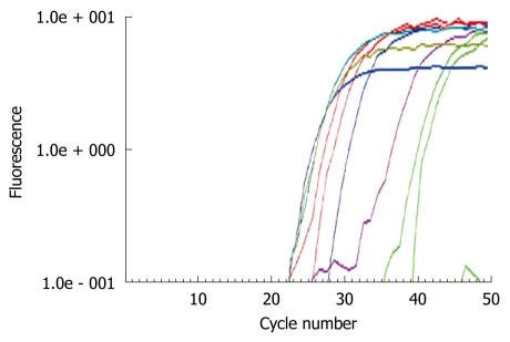
Methods: Expression of GPC3, Wnt, matrix metalloproteinases (MMPs), sulfatase (SULF)1, SULF2, and other growth signaling molecules was analyzed in HCC cell lines and tissue samples by real-time reverse transcription-polymerase chain reaction, immunoblotting, and/or immunostaining. Expression of various genes in GPC3 siRNA-transfected HCC cells was analyzed.
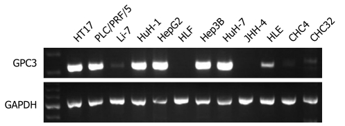
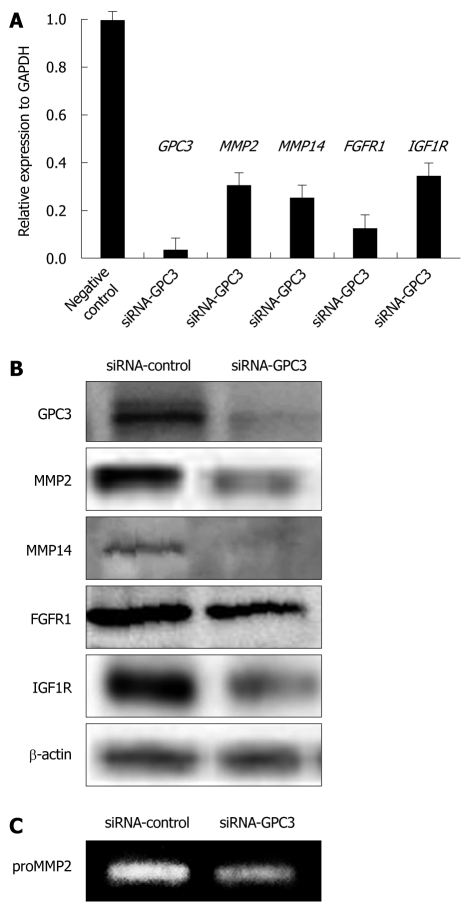
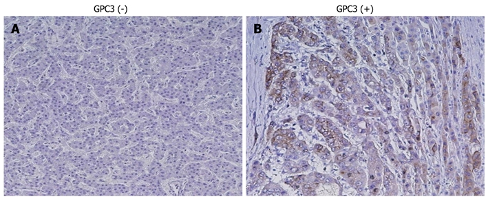
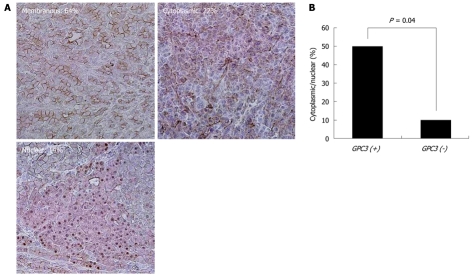
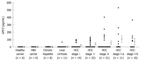
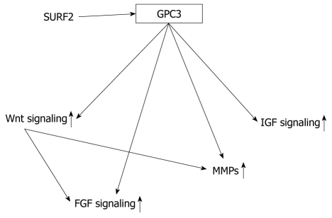
Results: GPC3 was overexpressed in most HCCs at mRNA and protein levels and its serum levels were significantly higher in patients with HCC than in non-HCC subjects (P < 0.05). Altered expressions of various MMPs and growth signaling molecules, some of which were correlated with GPC3 expression, were observed in HCCs. Down-regulation of GPC3 expression by siRNA in GPC3-overexpressing HCC cell lines resulted in a significant decrease in expressions of MMP2, MMP14, fibroblast growth factor receptor 1, insulin-like growth factor 1 receptor. GPC3 expression was significantly correlated with nuclear/cytoplasmic localization of beta-catenin.
Conclusion: These results suggest that GPC3, in conjunction with MMPs and growth signaling molecules, might play an important role in the progression of HCC.
Figures
Similar articles
-
Sulfatase 2 up-regulates glypican 3, promotes fibroblast growth factor signaling, and decreases survival in hepatocellular carcinoma.Hepatology. 2008 Apr;47(4):1211-22. doi: 10.1002/hep.22202. Hepatology. 2008. PMID: 18318435 Free PMC article.
-
The oncogenic effect of sulfatase 2 in human hepatocellular carcinoma is mediated in part by glypican 3-dependent Wnt activation.Hepatology. 2010 Nov;52(5):1680-9. doi: 10.1002/hep.23848. Hepatology. 2010. PMID: 20725905 Free PMC article.
-
Glypican-3 binds to Frizzled and plays a direct role in the stimulation of canonical Wnt signaling.J Cell Sci. 2014 Apr 1;127(Pt 7):1565-75. doi: 10.1242/jcs.140871. Epub 2014 Feb 4. J Cell Sci. 2014. PMID: 24496449
-
Glypican-3: a marker and a therapeutic target in hepatocellular carcinoma.FEBS J. 2013 May;280(10):2471-6. doi: 10.1111/febs.12126. Epub 2013 Jan 31. FEBS J. 2013. PMID: 23305321 Review.
-
Glypican-3: A promising biomarker for hepatocellular carcinoma diagnosis and treatment.Med Res Rev. 2018 Mar;38(2):741-767. doi: 10.1002/med.21455. Epub 2017 Jun 16. Med Res Rev. 2018. PMID: 28621802 Review.
Cited by
-
Current Trends in Non-Invasive Imaging of Interactions in the Liver Tumor Microenvironment Mediated by Tumor Metabolism.Cancers (Basel). 2021 Jul 21;13(15):3645. doi: 10.3390/cancers13153645. Cancers (Basel). 2021. PMID: 34359547 Free PMC article. Review.
-
Glypican-3 Expression in Patients with Oral Squamous Cell Carcinoma.J Dent (Shiraz). 2020 Jun;21(2):141-146. doi: 10.30476/DENTJODS.2019.84541.1089. J Dent (Shiraz). 2020. PMID: 32582830 Free PMC article.
-
An Atlas of Heparan Sulfate Proteoglycans in the Postnatal Rat Lens.Invest Ophthalmol Vis Sci. 2021 Nov 1;62(14):5. doi: 10.1167/iovs.62.14.5. Invest Ophthalmol Vis Sci. 2021. PMID: 34730792 Free PMC article.
-
The Role of Small Interfering RNAs in Hepatocellular Carcinoma.J Gastrointest Cancer. 2024 Mar;55(1):26-40. doi: 10.1007/s12029-023-00911-w. Epub 2023 Jul 11. J Gastrointest Cancer. 2024. PMID: 37432548 Review.
-
Glypican-3 deficiency in liver cancer upregulates MAPK/ERK pathway but decreases cell proliferation.Am J Cancer Res. 2024 Jul 15;14(7):3348-3371. doi: 10.62347/TTNY4279. eCollection 2024. Am J Cancer Res. 2024. PMID: 39113871 Free PMC article.
References
-
- El-Serag HB, Rudolph KL. Hepatocellular carcinoma: epidemiology and molecular carcinogenesis. Gastroenterology. 2007;132:2557–2576. - PubMed
-
- Llovet JM, Burroughs A, Bruix J. Hepatocellular carcinoma. Lancet. 2003;362:1907–1917. - PubMed
-
- Takagi H, Sasaki S, Suzuki H, Toyota M, Maruyama R, Nojima M, Yamamoto H, Omata M, Tokino T, Imai K, et al. Frequent epigenetic inactivation of SFRP genes in hepatocellular carcinoma. J Gastroenterol. 2008;43:378–389. - PubMed
-
- Hsu HC, Tseng HJ, Lai PL, Lee PH, Peng SY. Expression of p53 gene in 184 unifocal hepatocellular carcinomas: association with tumor growth and invasiveness. Cancer Res. 1993;53:4691–4694. - PubMed
Publication types
MeSH terms
Substances
LinkOut - more resources
Full Text Sources
Other Literature Sources
Medical
Research Materials
Miscellaneous
