

Role of Toll-like receptors in host responses to a virulence antigen of Streptococcus mutans expressed by a recombinant, attenuated Salmonella vector vaccine
- PMID: 20653102
- PMCID: PMC2910446
- DOI: 10.1016/j.vaccine.2010.05.039
Role of Toll-like receptors in host responses to a virulence antigen of Streptococcus mutans expressed by a recombinant, attenuated Salmonella vector vaccine
Abstract
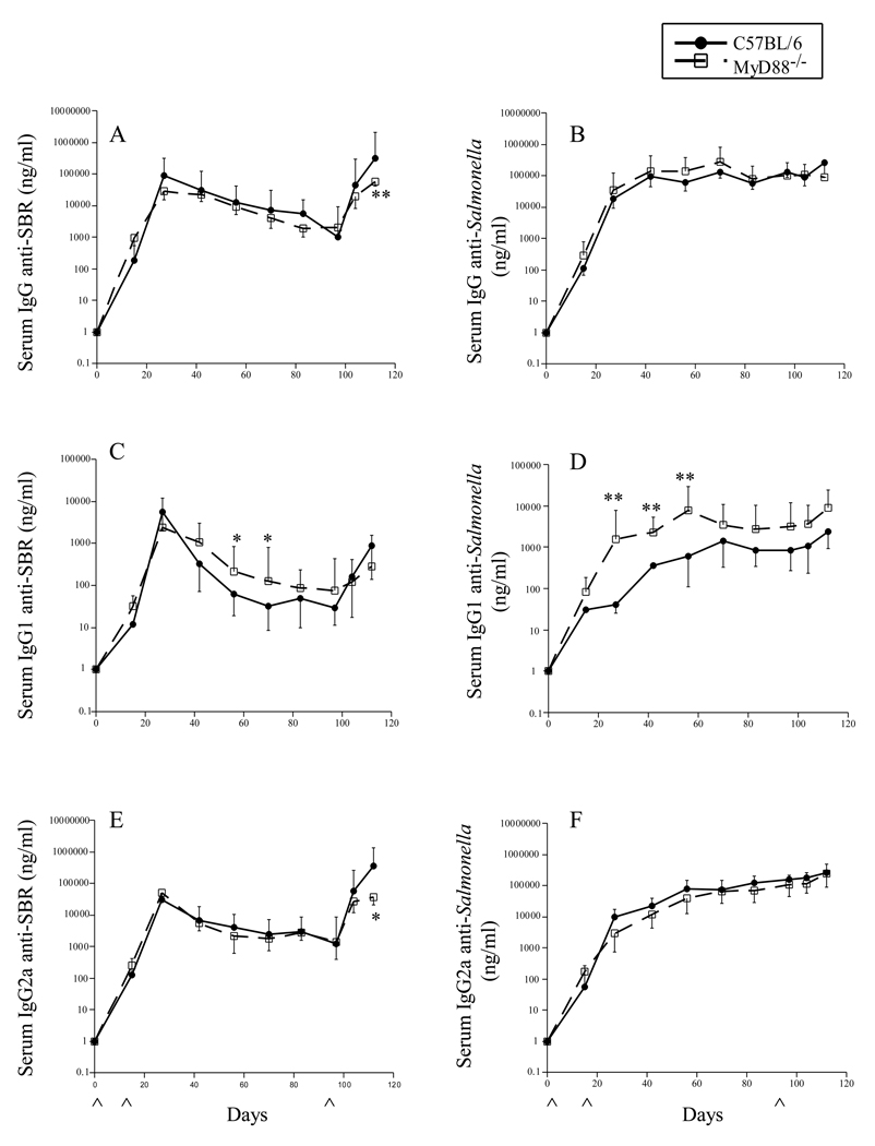
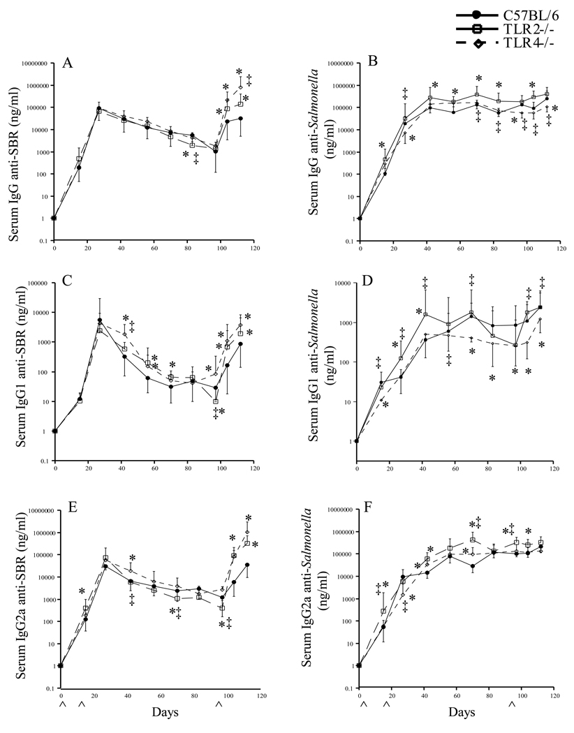
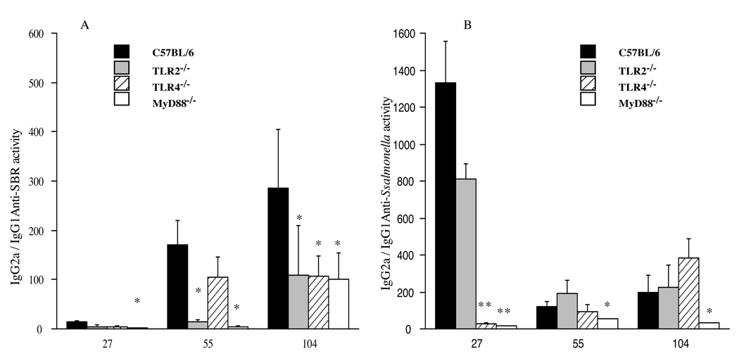
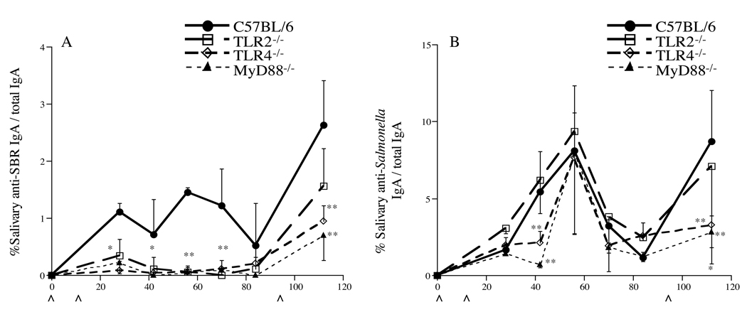
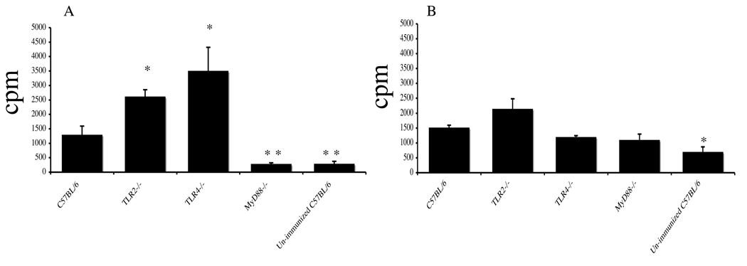
In the present study, we investigated the role of Toll-like receptors (TLRs) in host responses to the saliva-binding region (SBR) of Streptococcus mutans expressed by a recombinant, attenuated Salmonella vaccine. C57BL/6 wild type (wt), TLR2-/-, TLR4-/- and MyD88-/- mice were immunized by the intranasal route on days 0, 18 and boosted on day 98 with Salmonella typhimurium BRD 509 containing a plasmid encoding SBR. Serum and saliva samples were collected throughout the experiment and assessed for antibody activity by ELISA. Evidence is provided that the induction of a serum IgG2a (Th1-type) anti-SBR antibody response involved TLR2 signaling, whereas the anti-Salmonella response involved signaling through TLR4. The adaptor molecule MyD88 was not essential for the induction of a primary Th1-type response to SBR or Salmonella, but was necessary for a secondary response to SBR. Furthermore, the absence of TLR2, TLR4 or MyD88 resulted in enhanced Th2-type serum IgG1 anti-SBR and anti-Salmonella responses. Mucosal IgA responses to SBR were TLR2-, TLR4- and MyD88-dependent, while IgA responses to Salmonella were TLR4- and MyD88-dependent.
Figures
Similar articles
-
Contribution of a Streptococcus mutans antigen expressed by a Salmonella vector vaccine in dendritic cell activation.Infect Immun. 2011 Sep;79(9):3792-800. doi: 10.1128/IAI.05338-11. Epub 2011 Jul 11. Infect Immun. 2011. PMID: 21746857 Free PMC article.
-
Induction of protective immunity against Streptococcus mutans colonization after mucosal immunization with attenuated Salmonella enterica serovar typhimurium expressing an S. mutans adhesin under the control of in vivo-inducible nirB promoter.Infect Immun. 2001 Apr;69(4):2154-61. doi: 10.1128/IAI.69.4.2154-2161.2001. Infect Immun. 2001. PMID: 11254570 Free PMC article.
-
Enhanced immunogenicity of a genetic chimeric protein consisting of two virulence antigens of Streptococcus mutans and protection against infection.Infect Immun. 2002 Dec;70(12):6779-87. doi: 10.1128/IAI.70.12.6779-6787.2002. Infect Immun. 2002. PMID: 12438353 Free PMC article.
-
Innate immunity mediated by MyD88 signal is not essential for induction of lipopolysaccharide-specific B cell responses but is indispensable for protection against Salmonella enterica serovar Typhimurium infection.J Immunol. 2009 Feb 15;182(4):2305-12. doi: 10.4049/jimmunol.0801980. J Immunol. 2009. PMID: 19201885 Free PMC article.
-
Toll-like receptors and fungal infections: the role of TLR2, TLR4 and MyD88 in paracoccidioidomycosis.FEMS Immunol Med Microbiol. 2008 Jun;53(1):1-7. doi: 10.1111/j.1574-695X.2008.00378.x. Epub 2008 Apr 1. FEMS Immunol Med Microbiol. 2008. PMID: 18384366 Review.
Cited by
-
Contribution of a Streptococcus mutans antigen expressed by a Salmonella vector vaccine in dendritic cell activation.Infect Immun. 2011 Sep;79(9):3792-800. doi: 10.1128/IAI.05338-11. Epub 2011 Jul 11. Infect Immun. 2011. PMID: 21746857 Free PMC article.
-
Phosphate groups of lipid A are essential for Salmonella enterica serovar Typhimurium virulence and affect innate and adaptive immunity.Infect Immun. 2012 Sep;80(9):3215-24. doi: 10.1128/IAI.00123-12. Epub 2012 Jul 2. Infect Immun. 2012. PMID: 22753374 Free PMC article.
References
-
- Hajishengallis G, Michalek SM. Current status of a mucosal vaccine against dental caries. Oral Microbiol Immunol. 1999;14:1–20. - PubMed
-
- Russell MW, Lehner T. Characterization of antigens extracted from cells and culture fluids of Streptococcus mutans serotype c. Archs Oral Biol. 1978;23:7–15. - PubMed
-
- Hajishengallis G, Koga T, Russell MW. Affinity and specificity of the interactions between Streptococcus mutans antigen I/II and salivary components. J Dent Res. 1994;73:1493–1502. - PubMed
Publication types
MeSH terms
Substances
Grants and funding
LinkOut - more resources
Full Text Sources
Miscellaneous
