

Review
doi: 10.3109/10409238.2010.502516.
Epub 2010 Jul 23.
The ESCRT complexes
Affiliations
- PMID: 20653365
- PMCID: PMC2988974
- DOI: 10.3109/10409238.2010.502516
Item in Clipboard
Review
The ESCRT complexes
Crit Rev Biochem Mol Biol.
2010 Dec.
Abstract
The ESCRT machinery consists of the peripheral membrane protein complexes ESCRT-0, -I, -II, -III, and Vps4-Vta1, and the ALIX homodimer. The ESCRT system is required for degradation of unneeded or dangerous plasma membrane proteins; biogenesis of the lysosome and the yeast vacuole; the budding of most membrane enveloped viruses; the membrane abscission step in cytokinesis; macroautophagy; and several other processes. From their initial discovery in 2001-2002, the literature on ESCRTs has grown exponentially. This review will describe the structure and function of the six complexes noted above and summarize current knowledge of their mechanistic roles in cellular pathways and in disease.
Figures

Multivesicular bodies. Color versions of all figures are available on line.

Major cellular functions of the ESCRTs.

Schematic of the organization of ESCRT-0. a) yeast and (b) human ESCRT-0 shown docked to a flat, cargo-bearing membrane.

Schematic of the organization of ESCRT-I and –II. (a) yeast and (b) human ESCRT-I and –II depicted as a supercomplex assembled at a membrane neck.

Speculative schematic of the organization of ESCRT-III. The assembly is depicted as a spiral on the basis of the EM images of (Hanson et al., 2008) and for simplicity. The actual assembly is more likely dome-shaped rather than flat (Fabrikant et al., 2009). The order of assembly of the first four subunits (beginning with Vps20 at the outside of the spiral), and the preponderance of Snf7 subunits shown is based on the observations of (Teis et al., 2008).

Schematic of the organization of Vps4-Vta1.

Schematic of the organization of Bro1.

Ubiquitin binding domains. Ubiquitin is shown in yellow with Ile44 shown with space-filling spheres. UBDs are shown in blue. The VHS domain complex is shown for the human STAM subunit of ESCRT-0 (pdb entry 3LDZ) but is representative of all of the yeast and human ESCRT VHS domains. The UIM is shown for yeast ESCRT-0 subunit Vps27 (1Q0W) and is probably also representative of other “single” or conventional UIMs in the yeast ESCRT-0 Hse1 subunit and the human ESCRT-0 STAM subunit (Fig. 3). The DUIM is shown for human ESCRT-0 subunit Hrs (2D3G). The UEV domain is shown for human ESCRT-I (1S1Q) and is also representative of the yeast UEV-ubiquitin complex. The structure of the yeast Mvb12 C-terminal UBD in complex with ubiquitin has not been reported. The structure of the NZF of Npl4 is shown (1Q5W), as a reasonable representation of the yeast ESCRT-II NZF2-ubiquitin interaction, as no structure is available for the latter. This domain is not present in human ESCRT-II. The structure of the GLUE domain of human ESCRT-II is shown (2HTH); the yeast GLUE domain does not bind ubiquitin.

Membrane binding domains. Protein surfaces are colored green (hydrophobic residues), white (uncharged polar residues), red (acidic residues), and blue (basic residues). Structures are oriented such that the plane of the membrane is beneath each structure. The FYVE domain of ESCRT-0 (1VFY) targets phosphatidylinositol 3-phosphate, shown here as the soluble headgroup inositol 1,3-bisphosphate docked onto to the unliganded Vps27 FYVE domain structure from the liganded EEA1 FYVE structure (1JOC). The GLUE domain of yeast ESCRT-II (2CAY) is shown, with the non-canonical phosphoinositide binding pocket marked by a bound sulfate ion. The α1- α 4 membrane binding core of the ESCRT-III subunit VPS24 (2GD5) is shown, with the autoinhibitory helix omitted.

MIT domain-MIM complexes. The MIT domain of yeast Vps4(cyan) is shown bound to the MIM1 of Vps2 (2V6X). The MIT domain of human VPS4A (cyan) is shown in complex with the MIM2 of human VPS20 (2K3W). The MIT domain of human spastin (blue) is shown bound to the extended MIM1 of human DID2B (3EAB). MIMs are orange.

The Bro1 domain. The structure of the Bro1 domain of ALIX (cyan) is shown in complex with SNF7B (orange, 3C3Q). Conserved interaction residues are highlighted in stick models, and the Src binding site is also shown.
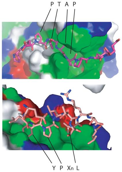
Viral late domains and ESCRTs. Top, the PTAP motif (stick model) of HIV-1 bound to the UEV domain of TSG101 (1M4Q). Botton, the YPX3L motif (stick model) of HIV-1 bound to the V domain of ALIX (2R02). Protein surfaces are colored green (hydrophobic residues), white (uncharged polar residues), red (acidic residues), and blue (basic residues).

GPPX3Y motif targeting to midbodies. The two subunits of CEP55 are colored cyan and orange, and the GPPX3Y peptide of ALIX is shown in a stick model.
References
-
- Agromayor M, Martin-Serrano J. Interaction of AMSH with ESCRT-III and deubiquitination of endosomal cargo. J Biol Chem. 2006;281:23083–23091. - PubMed
-
- Alam SL, Langelier C, Whitby FG, Koirala S, Robinson H, Hill CP, Sundquist WI. Structural basis for ubiquitin recognition by the human ESCRT-II EAP45 GLUE domain. Nat Struct Mol Biol. 2006;13:1029–1030. - PubMed
-
- Alam SL, Sundquist WI. Structural biology - ESCRT service. Nature. 2007;447:921–922. - PubMed
Publication types
MeSH terms
Substances
Grants and funding
LinkOut - more resources
Full Text Sources
Molecular Biology Databases
Miscellaneous