

Comparative analysis of total body and dermatoscopic photographic monitoring of nevi in similar patient populations at risk for cutaneous melanoma
- PMID: 20653722
- PMCID: PMC3025478
- DOI: 10.1111/j.1524-4725.2010.01589.x
Comparative analysis of total body and dermatoscopic photographic monitoring of nevi in similar patient populations at risk for cutaneous melanoma
Abstract
Background: Our previous experience monitoring nevi in high-risk patients using serial digital epiluminescence microscopy (DELM) photography achieved low biopsy rates but was limited by melanomas presenting as new lesions or arising from nevi that had not been photographed.
Objective: To determine whether biopsy rates, efficiency of melanoma detection, and melanoma origin (de novo vs nevus derived) differed in a similar patient population monitored using total body (TB) photography.
Methods: One thousand seventy-six patients (including 187 from a prior cohort) underwent TB photography and were monitored using photographs obtained at the initial visit. Risk factors and median monitoring periods for these patients were comparable with those of patients previously monitored using DELM photography.
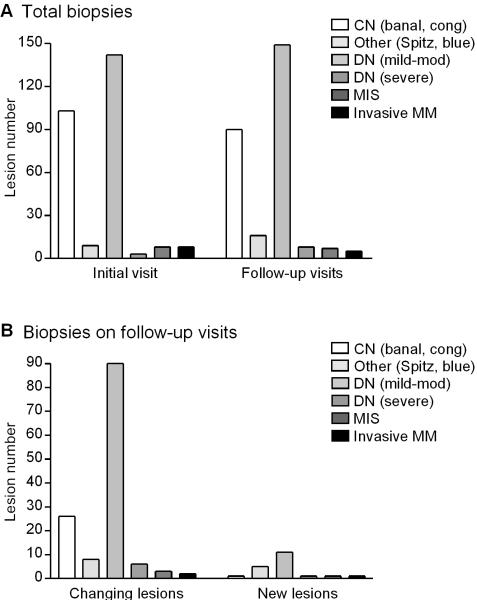
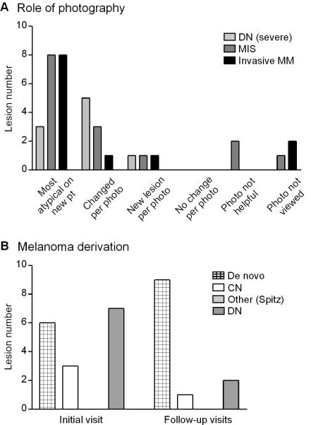
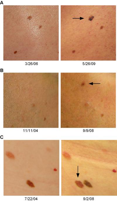
Results: Two hundred seventy-five biopsies were performed in 467 patients on follow-up visits. Of 12 melanomas detected on follow-up, five were invasive, five presented as changing lesions and two as new lesions, nine arose de novo, and the remainder were nevus derived.
Conclusions: In our experience with both approaches, monitoring patients at risk for melanoma using TB photography was associated with lower biopsy rates and lower nevus-to-melanoma ratios than using DELM and facilitated detection of new and changing lesions. In both cohorts, the majority of melanomas detected on follow-up arose de novo.
Figures
Comment in
-
Total body photography versus digital dermoscopic follow-up in the diagnosis of pigmented lesions.Dermatol Surg. 2011 Mar;37(3):406-7. doi: 10.1111/j.1524-4725.2011.01901.x. Dermatol Surg. 2011. PMID: 21410826 No abstract available.
References
-
- Tucker MA, et al. Risk of melanoma and other cancers in melanoma-prone families. J Invest Dermatol. 1993;100:350S–55S. - PubMed
-
- Kelly JW, et al. A high incidence of melanoma found in patients with multiple dysplastic naevi by photographic surveillance. Med J Aust. 1997;167:191–94. - PubMed
-
- Lucas CR, et al. Early melanoma detection: nonuniform dermoscopic features and growth. J Am Acad Dermatol. 2003;48:663–71. - PubMed
Publication types
MeSH terms
Grants and funding
LinkOut - more resources
Full Text Sources
Other Literature Sources
Medical
