

doi: 10.1111/j.1538-7836.2010.03995.x.
The generation and characterization of mice expressing a plasmin-inactivating active site mutation
- PMID: 20653841
- PMCID: PMC2965814
- DOI: 10.1111/j.1538-7836.2010.03995.x
Item in Clipboard
The generation and characterization of mice expressing a plasmin-inactivating active site mutation
J Thromb Haemost.
2010 Oct.
No abstract available
Figures
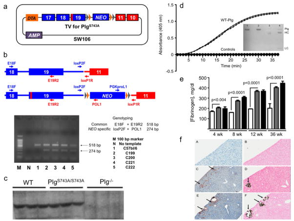
Generation and characterization of mice expressing an inactivating Plm active site mutation. (a). The final targeting vector for the PlgS743A mutation. Plg exons are in blue and Slc22a3 in red. The red vertical bar in Plg exon 19 is the location of the S743A mutation. The DTA autotoxic gene is present upstream of exon 17 such that undesired recombinants that possessed DTA would not survive. Lox (gray) and FRT (orange) sites flank the NEO gene in the 3’ UTR region of Plg. Positive selection of ES cells containing the NEO gene was made via kanamycin/ampicillin resistance. (b). PCR- and sequence-based identification of ES cells carrying the mutated allele. A common forward primer in Plg exon 18 (E18F) and a reverse primer in Plg exon 19 (E19R2) yields an amplicon of 518 bp for both WT Plg and PlgS743A. This amplified region was sequenced to confirm the S743A mutation. The primers loxP2F in the 3’ region of Plg and POL1 in the NEO gene provide a 274 bp amplicon only in the case of PlgS743A, and was sequenced to confirm the integrity of the 5’ lox and FRT sites. The amplicon generated by primers PGKproL1 and loxP1R was sequenced to confirm the identity of the 3’ FRT and lox sites. (c). Western blot analysis of plasmas from WT, PlgS743A/S743A, and Plg−/− mice using anti-mouse Plg chemiluminescence detection. (d). Activation of plasma Plg by uPA. Purified WT and PlgS743A/S743A (20 μg/ml) were incubated with 0.25 mM S2251 at 37°C. The activation of Plg was accelerated with 100 IU (relative to the WHO International Standard of high-molecular weight human uPA)/ml of recombinant high-molecular weight mouse uPA. Plm generation was continually monitored at 405 nm through hydrolysis of S2251 with liberation of p-nitroanilide. Control reactions, which are indistinguishable from each other in the Figure panel, consisted of WT Plg and PlgS743A/S743A without uPA, uPA without Plg, and S2251 without proteins. Plm activity was only detected in the duplicate samples of WT Plg with uPA. Insets are reduced SDS gels of purified plasma WT Plg (1) and PlgS743A/S743A (2), and WT Plg activated with uPA(3) and PlgS743A/S743A activated with uPA (4). Plm heavy (HC) and light (LC) chains are noted upon uPA treatment of WT Plg and PlgS743A/S743A in lanes 3 and 4. However, since active Plm is formed upon WT Plg activation, a doublet heavy chain consisting of Glu1-HC and Lys78-HC of Plm is seen. Active Plm is not formed upon activation of PlgS743A/S743A and only the Glu1-HC of Plm is observed. (e). Fibrinogen levels in resting WT and PlgS743A/S743A mice between 4–36 wk of age. The white, gray, and black bars represent WT Pg, PlgS743A/S743A, and Plm−/−mice, respectively at the various ages. N = 6–10 mice or each genotype at each age. (f). Histological stains of liver slices (4 μm) from WT (A, B). Plg−/− (C, D), and PlgS743A/S743A (E, F) mice for anti-fibrin(ogen) (A, C, E) immunostaining and von Kossa silver staining for calcium (B, D, F), scanned at 20X. The arrows indicate examples of the positive areas of the slides.
References
-
- Castellino FJ, Ploplis VA. Plasminogen Structure, activation, and regulation. Kluwer Academic/Plenum Publishers; 2003. Human plasminogen: structure, activation, and function; pp. 3–17.
-
- Sodeinde OA, Subrahmanyam YV, Stark K, Quan T, Bao Y, Goguen JD. A surface protease and the invasive character of plague. Science. 1992;258:1004–7. - PubMed
-
- Schott D, Dempfle CE, Beck P, Liermann A, Mohr-Pennert A, Goldner M, Mehlem P, Azuma H, Schuster V, Mingers AM, Schwarz HP, Kramer MD. Therapy with a purified plasminogen concentrate in an infant with ligneous conjunctivitis and homozygous plasminogen deficiency. N Engl J Med. 1998;339:1679–86. - PubMed
-
- Drew AF, Kaufman AH, Kombrinck KW, Danton MJS, Daugherty CC, Degen JL, Bugge TH. Ligneous conjunctivitis in plasminogen-deficient mice. Blood. 1998;91:1616–24. - PubMed
Publication types
MeSH terms
Substances
Grants and funding
LinkOut - more resources
Full Text Sources
Molecular Biology Databases
