. 2010 Jan;12(1):1-12.
doi: 10.1111/j.1438-8677.2009.00206.x.

Dismantling of Arabidopsis thaliana mesophyll cell chloroplasts during natural leaf senescence

Affiliations

Dismantling of Arabidopsis thaliana mesophyll cell chloroplasts during natural leaf senescence

I M Evans et al. Plant Biol (Stuttg). 2010 Jan.

Abstract

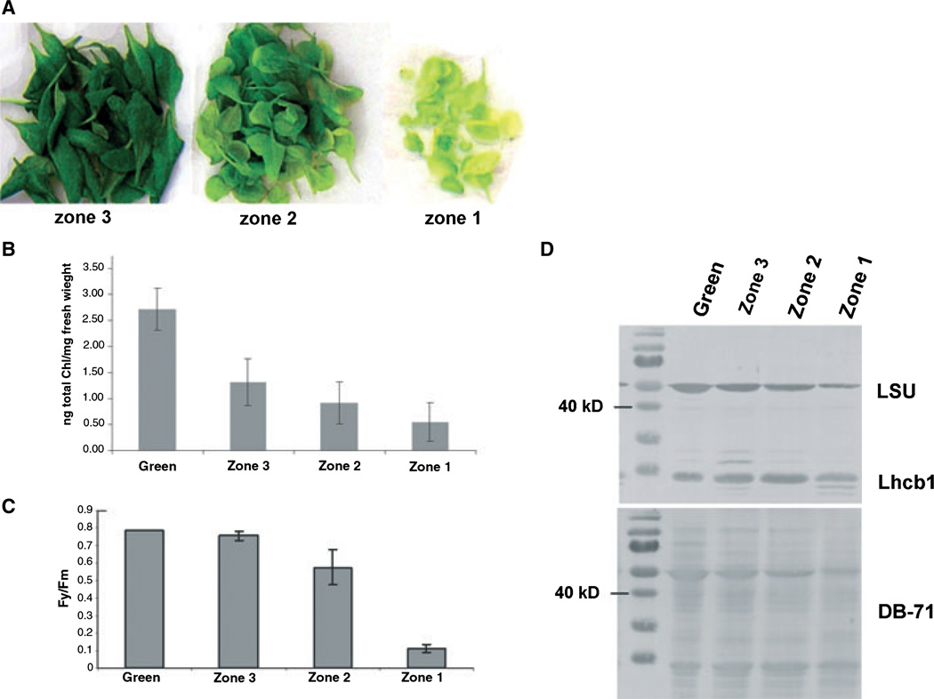
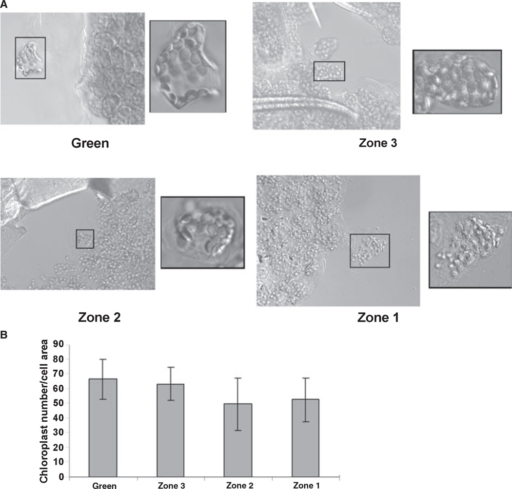
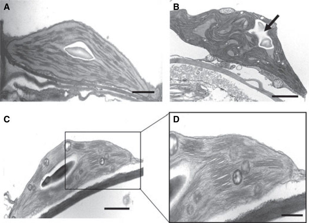
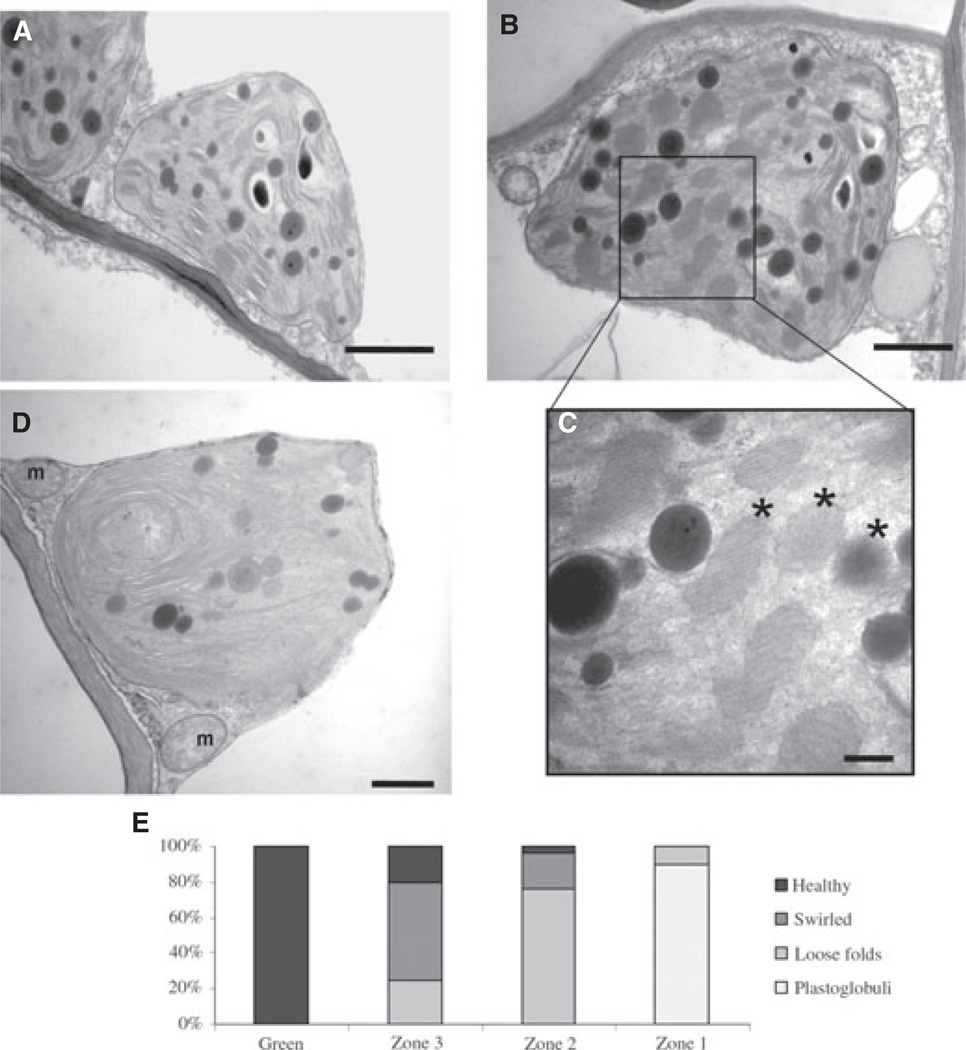
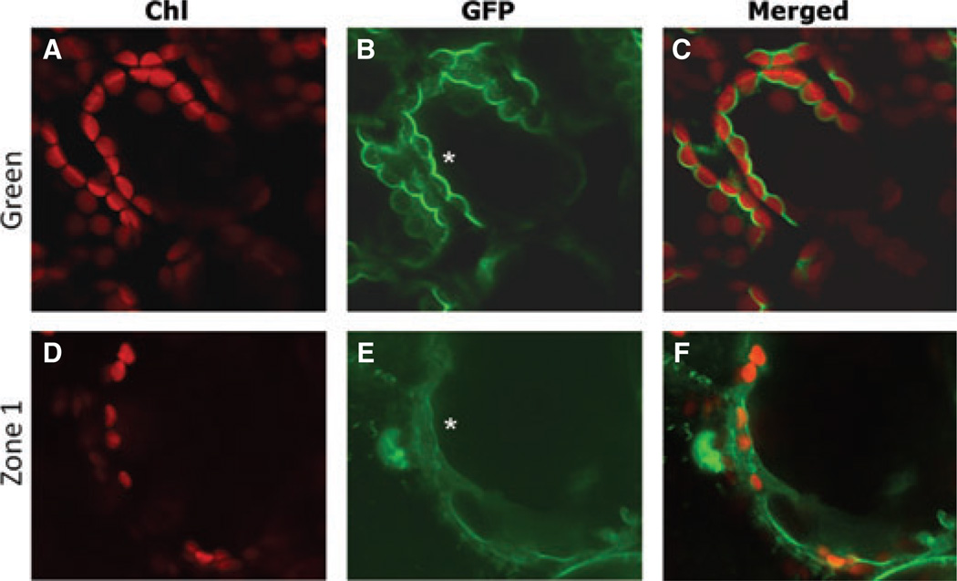
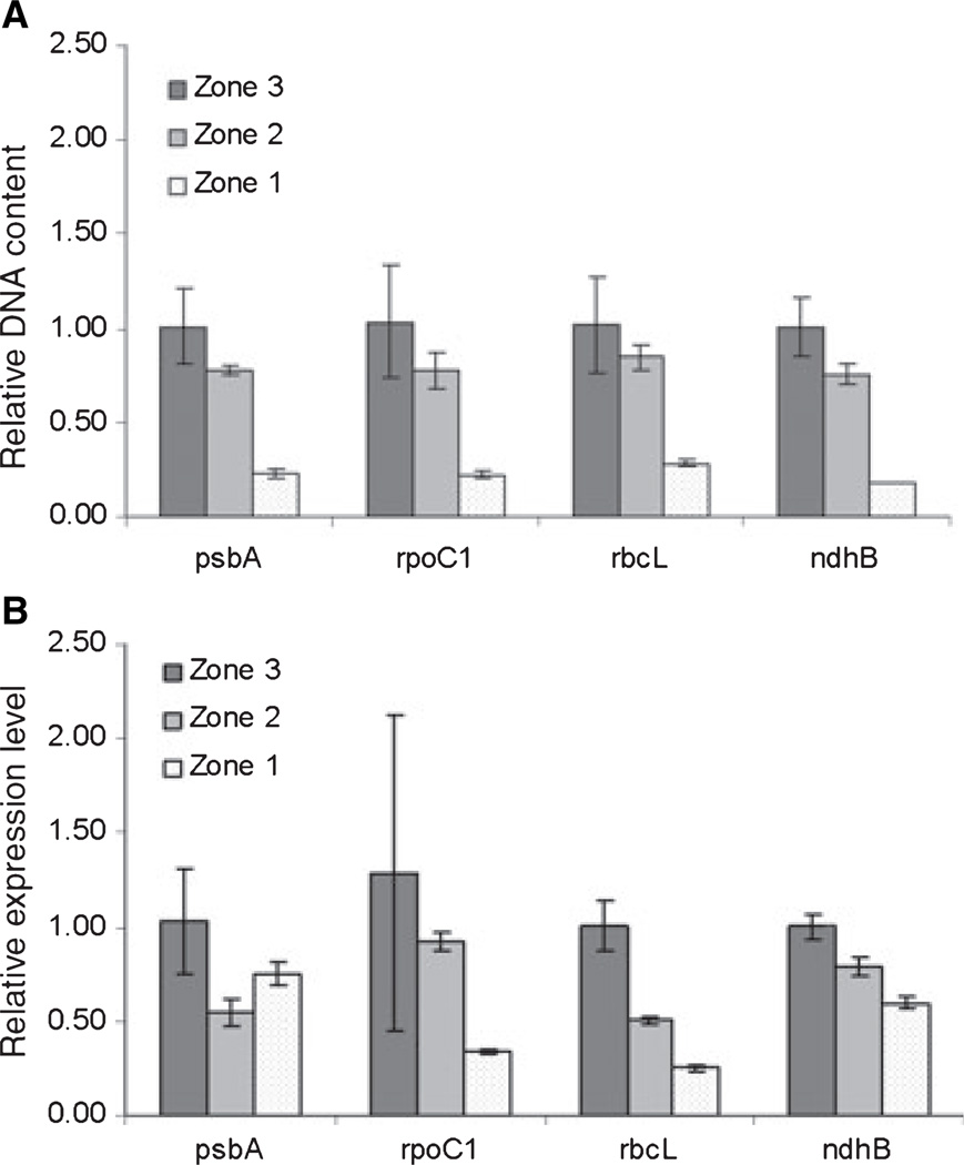
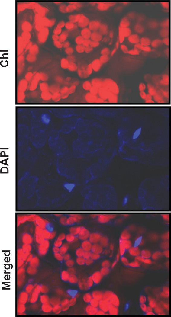
One of the earliest events in the process of leaf senescence is dismantling of chloroplasts. Mesophyll cell chloroplasts from rosette leaves were studied in Arabidopsis thaliana undergoing natural senescence. The number of chloroplasts decreased by only 17% in fully yellow leaves, and chloroplasts were found to undergo progressive photosynthetic and ultrastructural changes as senescence proceeded. In ultrastructural studies, an intact tonoplast could not be visualized, thus, a 35S-GFP::delta-TIP line with a GFP-labeled tonoplast was used to demonstrate that chloroplasts remain outside of the tonoplast even at late stages of senescence. Chloroplast DNA was measured by real-time PCR at four different chloroplast loci, and a fourfold decrease in chloroplast DNA per chloroplast was noted in yellow senescent leaves when compared to green leaves from plants of the same age. Although chloroplast DNA did decrease, the chloroplast/nuclear gene copy ratio was still 31:1 in yellow leaves. Interestingly, mRNA levels for the four loci differed: psbA and ndhB mRNAs remained abundant late into senescence, while rpoC1 and rbcL mRNAs decreased in parallel to chloroplast DNA. Together, these data demonstrate that, during senescence, chloroplasts remain outside of the vacuole as distinct organelles while the thylakoid membranes are dismantled internally. As thylakoids were dismantled, Rubisco large subunit, Lhcb1, and chloroplast DNA levels declined, but variable levels of mRNA persisted.


Figures

Fig. 1
Fig. 1
Zoning system for leaf senescence in Arabidopsis. A: Leaves were obtained from 7- to 8-week-old Arabidopsis rosettes, and separated into zones by visual inspection. Green tissue was harvested from mature leaves of 4-week-old plants while Zones 1–3 were harvested from 7- to 8-week-old plants. B: Total Chl was extracted from leaves (n = 8), and the average levels of Chl normalized to fresh weight ± SD are shown. C: Photochemical efficiency of photosytem II (Fv/Fm) was measured (n = 3), and the average values ± SD are shown. D: The levels of Rubisco LSU and Lhcb1 in each zone are shown in this immunoblot. Equal amounts of protein were loaded, as shown by DB-71 staining of the nitrocellulose filter prior to reaction with antibodies.
Fig. 2
Fig. 2
Chloroplast count per cell area. A: Mesophyll tissue was fixed, gently macerated, and then viewed by differential interference contrast microscopy. Chloroplasts were found to be plentiful in all zones observed. Higher magnification insets show chloroplasts at the surface of mesophyll cells. B: Chloroplasts were counted and then normalized to cell area (n = 16). Average values ± SD are shown.
Fig. 3
Fig. 3
TEM micrographs of green, Zone 3, and Zone 2 tissues. A: A chloroplast from a green section has normal thylakoid membranes and a starch granule in the center; bar = 500 nm. B: A section from Zone 3 tissue showing swirling thylakoid membranes (top arrow) and a large starch granule; bar = 500 nm. C: Zone 2 tissue shows the coalescence of membranes into large, loose folds; bar = 500 nm. D: Higher magnification of (C); bar = 200 nm.
Fig. 4
Fig. 4
TEM micrographs of Zone 1 tissue and quantitation of chloroplast ultrastructure types in each zone. A: A chloroplast in Zone 1 has plastoglobuli and membrane folds; bar = 1000 nm. B: Circular membrane folds can be seen; bar = 200 nm. C: Higher magnification of (B) shows the loss of membrane folds and formation of more densely staining plastoglobuli, indicated by asterisks; bar = 100 nm. D: Intact, late-senescent chloroplast; mitochondria (m) are present and inner membranes appear normal; bar = 200 nm. E: Fifty chloroplasts from each zone were assigned an ultrastructure type, and tallies were plotted.
Fig. 5
Fig. 5
The 35S-GFP::δ-TIP line observed by confocal microscopy. Chl autofluorescence is observed in panels (A) and (D), GFP fluorescence is observed in panels (B) and (E), and the merged images can be seen in panels (C) and (F). Leaves from 4-week-old plants are shown in the top three panels, and yellow, Zone 1 leaves from 7-week-old plants are shown in the bottom three panels. Asterisks indicate regions where the tonoplast contours the chloroplasts.
Fig. 6
Fig. 6
Real-time qPCR of nuclear and chloroplast DNA and cDNA. A: Genomic DNA was isolated from the three zones, and subjected to real-time PCR analysis using four chloroplast genes and one nuclear gene (Table 1). The nuclear gene served as a reference to control for equal loading, while Zone 3 served as the calibrator for 2−ΔΔCT analysis. The fold change in DNA content compared to Zone 3 leaves is shown, along with standard deviation. CT values shown in Table 1 demonstrate that chloroplast DNA amplifies after far fewer PCR cycles than nuclear DNA. This experiment was repeated twice, and one representative experiment is shown. B: RNA isolated from each zone was copied into cDNA, and used as a template for real-time qPCR. This experiment was repeated three times, and one representative experiment is shown.
Fig. 7
Fig. 7
DAPI staining of mature Arabidopsis leaves. Leaves were fixed in glutaraldehyde and then stained with DAPI. The top panel shows Chl autofluorescence, the middle panel shows DAPI fluorescence, and the bottom panel is a merged image. Nuclei stained brightly with DAPI, while chloroplasts showed light staining, which was brightest at the periphery and outside of the chloroplast.

References

    1. Austin JR, Frost E, Vide P-A, Kessler F, Staehelin LA. Plastoglobules are lipoprotein subcompartments of the chloroplast that are permanently coupled to thylakoid membranes and contain biosynthetic enzymes. Plant Cell. 2006;18:1693–1703. - PMC - PubMed
    1. Avila EL, Zouhar J, Agee AE, Carter DG, Chary NS, Raikhel NV. Tools to study plant organelle biogenesis. Point mutation lines with disrupted vacuoles and high-speed confocal screening of green fluorescent protein-tagged organelles. Plant Physiology. 2003;133:1673–1676. - PMC - PubMed
    1. Barton R. Fine structure of mesophyll cells in senescing leaves of Phaseolus. Planta. 1966;71:314–325. - PubMed
    1. Buchanan-Wollaston V, Page T, Harrison E, Breeze E, Lim PO, Nam HG, Lin J-F, Wu S-H, Swidzinski J, Ishizaki K, Leaver CJ. Comparative transcriptome analysis reveals significant differences in gene expression and signalling pathways between developmental and dark / starvation- induced senescence in Arabidopsis. Plant Journal. 2005;42:567–585. - PubMed
    1. Chiba A, Ishida H, Nishizawa NK, Makino A, Mae T. Exclusion of ribulose-1,5-bisphosphate carboxylase / oxygenase from chloroplasts by specific bodies in naturally senescing leaves of wheat. Plant and Cell Physiology. 2003;44:914–921. - PubMed

Publication types

MeSH terms

LinkOut - more resources