. 2010 Jul 23;11(1):101.
doi: 10.1186/1465-9921-11-101.

Host lung gene expression patterns predict infectious etiology in a mouse model of pneumonia

Affiliations

Host lung gene expression patterns predict infectious etiology in a mouse model of pneumonia

Scott E Evans et al. Respir Res. .

Abstract

Background: Lower respiratory tract infections continue to exact unacceptable worldwide mortality, often because the infecting pathogen cannot be identified. The respiratory epithelia provide protection from pneumonias through organism-specific generation of antimicrobial products, offering potential insight into the identity of infecting pathogens. This study assesses the capacity of the host gene expression response to infection to predict the presence and identity of lower respiratory pathogens without reliance on culture data.

Methods: Mice were inhalationally challenged with S. pneumoniae, P. aeruginosa, A. fumigatus or saline prior to whole genome gene expression microarray analysis of their pulmonary parenchyma. Characteristic gene expression patterns for each condition were identified, allowing the derivation of prediction rules for each pathogen. After confirming the predictive capacity of gene expression data in blinded challenges, a computerized algorithm was devised to predict the infectious conditions of subsequent subjects.

Results: We observed robust, pathogen-specific gene expression patterns as early as 2 h after infection. Use of an algorithmic decision tree revealed 94.4% diagnostic accuracy when discerning the presence of bacterial infection. The model subsequently differentiated between bacterial pathogens with 71.4% accuracy and between non-bacterial conditions with 70.0% accuracy, both far exceeding the expected diagnostic yield of standard culture-based bronchoscopy with bronchoalveolar lavage.

Conclusions: These data substantiate the specificity of the pulmonary innate immune response and support the feasibility of a gene expression-based clinical tool for pneumonia diagnosis.


Figures

Figure 1
Figure 1
Survival following infectious challenges. (A) Using an experimental model of inhalational pneumonia in BALB/c mice, P. aeruginosa and S. pneumoniae both induced consistent mortality >80%, while mice challenged with A. fumigatus or PBS (sham) had 100% survival. (B) Mice treated with cyclophosphamide and cortisol prior to infection also consistently succumbed to A. fumigatus challenge, substantiating the effective delivery of pathogens to the mice (N = 10 mice/group, *p = 0.0007 vs. A. fumigatus, **p = 0.0001 vs. A. fumigatus, †p < 0.0001 vs. A. fumigatus).
Figure 2
Figure 2
Proteomic analysis of post-challenge BAL fluid. Mice were challenged with aerosolized P. aeruginosa, S. pneumoniae, A. fumigatus or PBS (sham). 24 h later, BAL was performed and concentrations of 16 cytokines and chemokines were measured by ELISA. In all cases, P. aeruginosa induced the highest level of cytokine or chemokine expression, with no test identifying any other infectious condition. Shown are representative examples: (A) Interferon-γ, (B) tumor necrosis factor (C) Interleukin-6 and (D) CCL17. (mean ± SD, N = 5 mice/group, *p < 0.005 compared to all other conditions.
Figure 3
Figure 3
Early development of infection-specific transcription profiles. (A) Six hours after challenge with P. aeruginosa, S. pneumoniae, A. fumigatus or PBS (sham), lungs were removed and submitted to microarray analysis, and a heatmap was generated with green indicating decreased gene expression and red indicating increased gene expression. At this time, 4,274 genes were highly differentially expressed (FDR< 1 × 10⁻⁷), and by unsupervised clustering, most samples self-segregated by challenge. (N = 6 sham infected mice, 8 mice for each infection.) (B) The 30 genes that were most strongly differentially expressed at 18 h after infection were examined at earlier time points, demonstrating the increasing clarity of the differential pattern. (N = 6 sham infected mice, 4 mice for each infection.)
Figure 4
Figure 4
Differential gene expression 18 hours after infectious challenge. (A) A heatmap shows the expression patterns of 367 DEGs after inhalational challenge with P. aeruginosa, S. pneumoniae, A. fumigatus or PBS (sham). By unsupervised clustering, the samples all correctly segregate themselves by condition. (B) A Venn diagram indicates the striking specificity of these expression patterns, with <10% of DEGs induced or repressed by more than one condition.
Figure 5
Figure 5
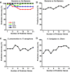
Diagnostic accuracy of computerized gene expression interrogation. Mice were exposed to one of four potential infectious conditions, then gene expression profiling was performed at designated time points after the challenge. (A) Diagnostic accuracy of algorithmic predictions of whether or not different mice were infected with bacteria, based on the time after infection and the number of transcripts used in the prediction model. (B-D) Rules derived from initial 18 h experiments were used to predict the infectious conditions of different mice 18 h after challenge in a separate validation set, based on number of transcripts in the algorithm. (B) Prediction accuracy for discriminating bacteria vs. non-bacteria. (C) Prediction accuracy for discriminating S. pneumoniae infection from P. aeruginosa infection. (D) Prediction accuracy for discriminating A. fumigatus from sham infection.

References

    1. WHO. The World Health Report 2004 -- Changing History. Geneva: World Health Organization; 2004.
    1. Rano A, Agusti C, Jimenez P, Angrill J, Benito N, Danes C, Gonzalez J, Rovira M, Pumarola T, Moreno A. et al.Pulmonary infiltrates in non-HIV immunocompromised patients: a diagnostic approach using non-invasive and bronchoscopic procedures. Thorax. 2001;56(5):379–387. doi: 10.1136/thorax.56.5.379. - DOI - PMC - PubMed
    1. Shorr AF, Susla GM, O'Grady NP. Pulmonary infiltrates in the non-HIV-infected immunocompromised patient: etiologies, diagnostic strategies, and outcomes. Chest. 2004;125(1):260–271. doi: 10.1378/chest.125.1.260. - DOI - PubMed
    1. White P, Bonacum JT, Miller CB. Utility of fiberoptic bronchoscopy in bone marrow transplant patients. Bone Marrow Transplant. 1997;20(8):681–687. doi: 10.1038/sj.bmt.1700957. - DOI - PubMed
    1. Bissinger AL, Einsele H, Hamprecht K, Schumacher U, Kandolf R, Loeffler J, Aepinus C, Bock T, Jahn G, Hebart H. Infectious pulmonary complications after stem cell transplantation or chemotherapy: diagnostic yield of bronchoalveolar lavage. Diagn Microbiol Infect Dis. 2005;52(4):275–280. doi: 10.1016/j.diagmicrobio.2005.03.005. - DOI - PubMed

Publication types

MeSH terms