. 2010 Jul 23:10:76.
doi: 10.1186/1471-213X-10-76.

A novel transgenic zebrafish model for blood-brain and blood-retinal barrier development

Affiliations

A novel transgenic zebrafish model for blood-brain and blood-retinal barrier development

Jing Xie et al. BMC Dev Biol. .

Abstract

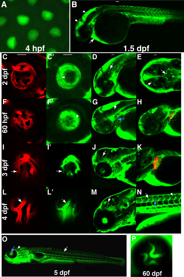
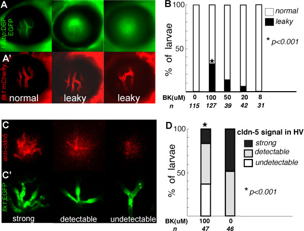
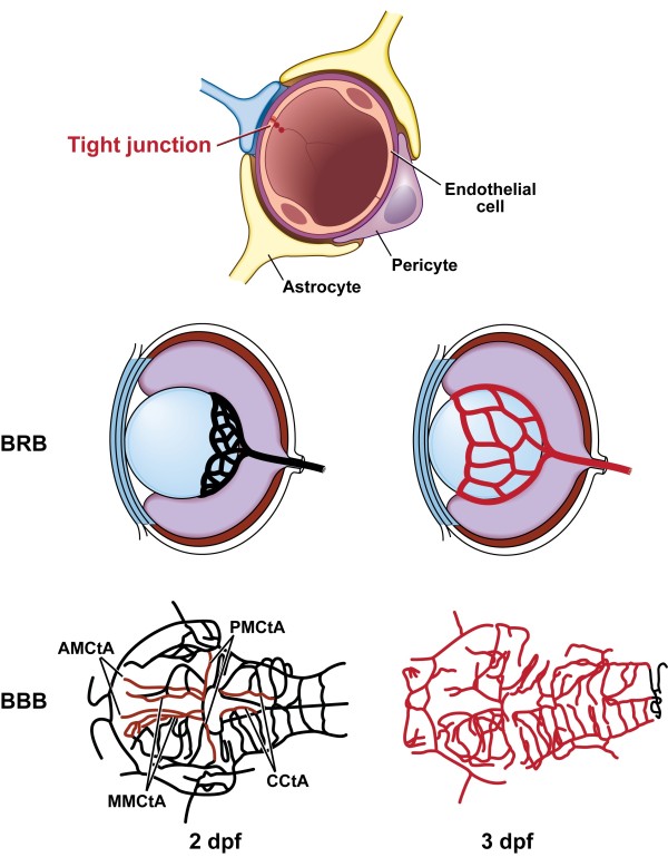
Background: Development and maintenance of the blood-brain and blood-retinal barrier is critical for the homeostasis of brain and retinal tissue. Despite decades of research our knowledge of the formation and maintenance of the blood-brain (BBB) and blood-retinal (BRB) barrier is very limited. We have established an in vivo model to study the development and maintenance of these barriers by generating a transgenic zebrafish line that expresses a vitamin D-binding protein fused with enhanced green fluorescent protein (DBP-EGFP) in blood plasma, as an endogenous tracer.

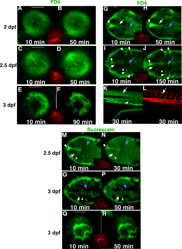
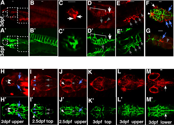
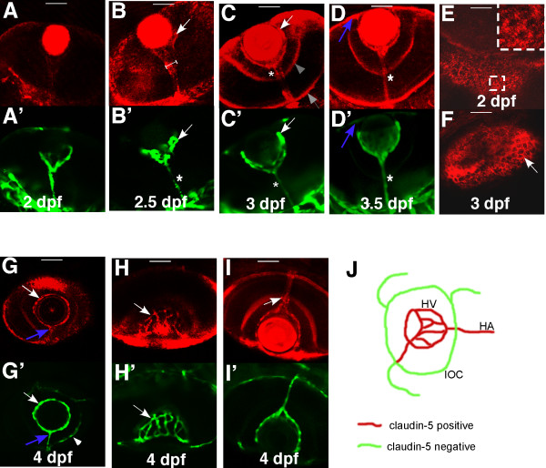
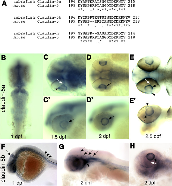
Results: The temporal establishment of the BBB and BRB was examined using this transgenic line and the results were compared with that obtained by injection of fluorescent dyes into the sinus venosus of embryos at various stages of development. We also examined the expression of claudin-5, a component of tight junctions during the first 4 days of development. We observed that the BBB of zebrafish starts to develop by 3 dpf, with expression of claudin-5 in the central arteries preceding it at 2 dpf. The hyaloid vasculature in the zebrafish retina develops a barrier function at 3 dpf, which endows the zebrafish with unique advantages for studying the BRB.

Conclusion: Zebrafish embryos develop BBB and BRB function simultaneously by 3 dpf, which is regulated by tight junction proteins. The Tg(l-fabp:DBP-EGFP) zebrafish will have great advantages in studying development and maintenance of the blood-neural barrier, which is a new application for the widely used vertebrate model.


Figures

Figure 1
Figure 1
Development of the BRB and BBB in zebrafish. FD4 (4,000 Da, green) was injected to the sinus venosus of Tg(flk1:mCherry) embryos at 2 dpf (A, B, G, H), 2.5 dpf (C, D, I, J) and 3 dpf (E, F, K. L). Live confocal images of the hyaloid vessels (A-F, side view), brain vessels (G-J, dorsal view) and trunk vessels (K-L, side view) were obtained from 10 to 150 minutes after injection. Insets and panel L are images of established vasculature of the injected embryos (red). In the brain, boundaries of the middle mesencephalic central artery (MMCtA) are indicated by arrows in G-J, cerebellar central artery (CCtA), by blue arrows in G-J and posterior mesencephalic central artery (PMCtA) by asterisks in I&J. Arrows in K&L indicate intersegmental vessels and arrowhead in K shows the myotomal boundaries. A small molecule, fluorescein (376 Da, green), was also used as a tracer in the injection assay (M-R). In the 2.5 dpf embryos (M&N), the PMCtA (asterisk) could not be differentiated, and boundaries of MMCtA (arrows) and CCtA (blue arrows) were enlarged and became blurred 30 minutes after injection. However in the 3 dpf embryos injected with fluorescein (O-R), boundaries of the MMCtA (arrows), PMCtA (asterisk), CCtA (blue arrows) and the hyaloid vessels (Q&R) remained sharp and clear after 50 minutes of injection. The leaked FD4 accumulated mostly in brain ventricles (arrowheads in G-J). In contrast, most of the leaked fluorescein did not accumulated in the brain ventricle (arrowheads in M&N), but evenly diffused throughout the brain (M-P). Scale bars: 50 um.
Figure 2
Figure 2
Spatial and temporal expression of claudin-5 in the developing BBB. Tg(flk1:EGFP) embryos at 2 dpf, 2.5 dpf, and 3 dpf were stained with a monoclonal claudin-5 antibody. Confocal images of whole mount embryos were analyzed for claudin-5 expression (red) (A-E and H-M; Alexa Fluor 568 labeled secondary antibody) in developing blood vessels (green) (A'-E' and H'-M'; EGFP labeled vascular endothelial cells; F&G, merged pictures). All the samples are oriented with anterior to the left. A-E and A'-E', lateral views; other panels, dorsal views. Scale bars: 50 um.
Figure 3
Figure 3
Spatial and temporal expression of claudin-5 in the developing BRB. Tg(flk1:EGFP) embryos and larvae from 2 dpf to 4 dpf were stained with mouse anti-claudin-5. Confocal images of whole mount were analyzed for claudin-5 (red) (A-I; Alexa Fluor 568) expression in hyaloid blood vessels (green) (A'-D' and G'-I'; EGFP). All the panels are dorsal views except G&G'. The claudin-5 signal in the hyaloid vessels at 2 dpf (A&A') is minimal. At 2.5 dpf (B&B'), and 3 dpf (C&C') the staining in the hyaloid vasculature is increased. Claudin-5 is also expressed in the hyaloid artery (asterisks) and outer limiting membrane of the retina (shaded arrow) and inner plexiform layer (shaded arrowhead). (D&D') At 3.5 dpf, the wider cone-shape staining is lost and the claudin-5 signal overlaps completely with the HA (asterisks). The vessel connecting the HV and the inner optic circle (IOC) also has a strong claudin-5 signal (blue arrows in D&D', G&G'). (E&F) In the retina, the claudin-5 signal does not clearly outline the polygonal RPE (arrow) until 3 dpf. The insert is an enlarged view of the dashed square. At 4 dpf (G-I), the HV (arrows in G&G', H&H') and the HA, but not the choroidal vasculature (such as the IOC, indicated by arrowhead in G'), express claudin-5. The claudin-5 is also expressed around the foramen (opening) through which the HA penetrates the retina (arrow in I). The panel J is a schematic illustrating expression of claudin-5 in optic vasculature of zebrafish. Scale bars: 50 um.
Figure 4
Figure 4
Claudin-5a and 5b expression in hyaloid vasculature. (A) The 3' ends of three zebrafish claudin genes have significant homology with the C-terminal of mouse claudin-5. Claudin-5a (zgc 85723; GenBank: NM_213274) and claudin-5b (zgc 103419; GenBank: NM_001006044) are mostly homologous to claudin-5a of Fugu rubripes, with 82% identical (plus 8% similar) and 75% identical (plus 14% similar) amino acid sequences, respectively. The zebrafish claudin-h (GenBank: NM_131767) is mostly homologous to claudin-3a of Fugu. (B-E') Whole mount in situ hybridization of claudin-5a expression. (B) Claudin-5a is expressed in the CNS (midbrain, hindbrain, ventricular zone, and epiphysis) at 1 dpf. (C-E & C'-E') From 1.5 to 2.5 dpf, claudin-5mRNA is detected in the hyaloid vasculature (black arrows), hyaloid artery (white arrows) and the cornea (arrow heads). (F-H) Claudin-5b is expressed in the entire vascular system at 1 dpf (arrow and arrowheads in F), and is confined to the blood vessels of the brain (arrows in G) and cardiovascular system (arrowhead in G) at 2 dpf. Similar to claudin-5a, expression of claudin-5b in the hyaloid vasculature (black arrow in H) and hyaloid artery (white arrow in H) is seen at 1.5 dpf and lasts till 3 dpf. B-E, dorsal view; C'-E', side view; F-H, side view.
Figure 5
Figure 5
Expression pattern of DBP-EGFP in the blood plasma of l-fabp:DBP-EGFP transgenic zebrafish. Images were obtained from Tg(l-fabp:DBP-EGFP;flk1:mCherry) fish to visualize DBP-EGFP (green) and endothelial cells lining blood vessels (red)(mCherry). The fish were oriented with anterior to the left. All panels are side views except E. (A) green fluorescence in eggs laid by female Tg(l-fabp:DBP-EGFP) fish. (B) DBP-EGFP expression at 1.5 dpf. (C, C', D & E) DBP-EGFP expression at 2 dpf. The central arteries (arrows) are the first distinguishable blood vessels. (F, F', G & H) DBP-EGFP expression at 60 hpf. The dashed rectangles in H and K are merged pictures of red and green channels. The branchial arches (blue arrow) can be differentiated at the time, but boundaries of HV are still not clear (arrowhead). At 3 dpf (I, I', J & K), the boundaries of the HV become sharp (arrows in I&I'). More vessels appear in the liver (dashed rectangle) and more brain vessels can be seen (arrows in G and J). At 4 dpf (L, L', M & N), the EGFP-infused hyaloid and brain vessels can be easily differentiated from the fluorescent background (arrows in L, L' and M), as can the intersegmental vessels (arrow in N) and the dorsal aorta (blue arrow). Accumulation of DBP-EGFP in the myotomal boundaries (arrowhead) (N) is observed from leakage out of the vasculature. At 5 dpf (O), the HV (blue arrow) and the trunk vessels (arrow) are distinguishable, but the brain vessels are not (arrowhead), due to the increased fluorescent background in the brain. (P) At 60 dpf, the HV is still distinguishable.
Figure 6
Figure 6
Bradykinin mediated disruption of zebrafish BRB (A, A' & B). The l-fabp:DBP-EGFP;flk1:mCherry double transgenic larvae were treated with 8 to 100 μM BK from 5 to 9 dpf. (A) DBP-EGFP (green) in hyaloid vessels of control (left panel), leaky hyaloid vessels (middle and right panel); (A') endothelial lining of blood vessels, mCherry (red). (B) At 9 dpf, the larvae were scored for the presence of leaky hyaloid vessels. Both Fisher's test and chi-square test indicate that the BK treatment results in significantly increased numbers of larvae showing GFP leakage compared with those treated with control buffer (P < 0.001). (C) Claudin-5 expression (red) was evaluated by whole mount immunohistochemistry in Tg(flk1:EGFP) larvae exposed to 100 μM BK and scored as strong expression (left panel), detectable (middle panel) and undetectable (right panel); (C') endothelial lining of blood vessels, flk1:EGFP (green). (D) Approximately 47 BK treated and 46 untreated flk1:EGFP larvae were evaluated for claudin-5 expression in the hyaloid vasculature.
Figure 7
Figure 7
Schematic illustration of endothelial tight junctions and claudin-5 expression in retinal and brain vasculature of zebrafish embryos at 2 dpf and 3 dpf. Four pairs of the central artery express claudin-5 at 2 dpf (red). The figure was adapted from the online data of Dr. Brant Weinstein's lab (zfish.nichd.nih.gov/zfatlas). AMCtA: anterior mesencephalic central artery; MMCtA: middle mesencephalic central artery; PMCtA: posterior mesencephalic central artery; CCtA: cerebellar central artery.

Similar articles

Cited by

References

    1. Abbott NJ, Ronnback L, Hansson E. Astrocyte-endothelial interactions at the blood-brain barrier. Nat Rev Neurosci. 2006;7:41–53. doi: 10.1038/nrn1824. - DOI - PubMed
    1. Neuwelt E, Abbott NJ, Abrey L, Banks WA, Blakley B, Davis T, Engelhardt B, Grammas P, Nedergaard M, Nutt J. Strategies to advance translational research into brain barriers. Lancet Neurol. 2008;7:84–96. doi: 10.1016/S1474-4422(07)70326-5. - DOI - PubMed
    1. Erickson KK, Sundstrom JM, Antonetti DA. Vascular permeability in ocular disease and the role of tight junctions. Angiogenesis. 2007;10:103–17. doi: 10.1007/s10456-007-9067-z. - DOI - PubMed
    1. Dejana E, Tournier-Lasserve E, Weinstein BM. The control of vascular integrity by endothelial cell junctions: molecular basis and pathological implications. Dev Cell. 2009;16:209–21. doi: 10.1016/j.devcel.2009.01.004. - DOI - PubMed
    1. Hawkins BT, Davis TP. The blood-brain barrier/neurovascular unit in health and disease. Pharmacol Rev. 2005;57:173–85. doi: 10.1124/pr.57.2.4. - DOI - PubMed

Publication types

MeSH terms