. 2010 Jul 24;17(1):60.
doi: 10.1186/1423-0127-17-60.

Molecular characterization of the PhoPQ-PmrD-PmrAB mediated pathway regulating polymyxin B resistance in Klebsiella pneumoniae CG43

Affiliations

Molecular characterization of the PhoPQ-PmrD-PmrAB mediated pathway regulating polymyxin B resistance in Klebsiella pneumoniae CG43

Hsin-Yao Cheng et al. J Biomed Sci. .

Abstract

Background: The cationic peptide antibiotic polymyxin has recently been reevaluated in the treatment of severe infections caused by gram negative bacteria.

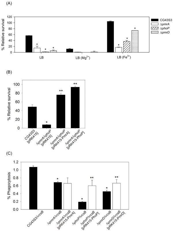
Methods: In this study, the genetic determinants for capsular polysaccharide level and lipopolysaccharide modification involved in polymyxin B resistance of the opportunistic pathogen Klebsiella pneumoniae were characterized. The expressional control of the genes responsible for the resistance was assessed by a LacZ reporter system. The PmrD connector-mediated regulation for the expression of pmr genes involved in polymyxin B resistance was also demonstrated by DNA EMSA, two-hybrid analysis and in vitro phosphor-transfer assay.

Results: Deletion of the rcsB, which encoded an activator for the production of capsular polysaccharide, had a minor effect on K. pneumoniae resistance to polymyxin B. On the other hand, deletion of ugd or pmrF gene resulted in a drastic reduction of the resistance. The polymyxin B resistance was shown to be regulated by the two-component response regulators PhoP and PmrA at low magnesium and high iron, respectively. Similar to the control identified in Salmonella, expression of pmrD in K. pneumoniae was dependent on PhoP, the activated PmrD would then bind to PmrA to prolong the phosphorylation state of the PmrA, and eventually turn on the expression of pmr for the resistance to polymyxin B.

Conclusions: The study reports a role of the capsular polysaccharide level and the pmr genes for K. pneumoniae resistance to polymyxin B. The PmrD connector-mediated pathway in governing the regulation of pmr expression was demonstrated. In comparison to the pmr regulation in Salmonella, PhoP in K. pneumoniae plays a major regulatory role in polymyxin B resistance.


Figures

Figure 1
Figure 1
Deletion effects of ugd, wza and rcsB genes on Klebsiella CPS production and resistance to polymyxin B. (A) Comparison of colony morphology. The K. pneumoniae strains were streaked on an LB agar plate, incubated at 37°C overnight and photographed. (B) Sedimentation test. The strains were cultured overnight in LB broth at 37°C and subjected to centrifugation at 4,000 ×g for 5 min. Quantification of K2 CPS amounts of each strain is shown below the figure. Values are shown as averages ± standard deviations from triplicate samples. (C) Polymyxin resistance assay. The log-phased cultures of K. pneumoniae CG43S3, Δugd, Δwza or ΔrcsB mutants were challenged with 1 or 2 units/ml of polymyxin B. (D) Polymyxin resistance assay. The log-phased culture of K. pneumoniae strains were challenged with 2 or 4 units/ml of polymyxin B. The survival rates are shown as the average ± standard deviations from triplicate samples. *, P < 0.01 compared to the parental strain CG43S3. **, P < 0.01 compared to each strain carrying pRK415.
Figure 2
Figure 2
Involvement of K. pneumoniae pmrF gene in polymyxin B resistance and intramacrophage survival. (A) The log-phased cultures of K. pneumoniae CG43S3, the ΔpmrF mutant or ΔpmrF carrying pRK415-PmrF were grown in LB or LB supplemented with 1 mM Fe3+ and then challenged with 2 units/ml of polymyxin B. The survival rates are shown as the average ± standard deviations from triplicate samples. (B) The survival rates of K. pneumoniae CG43S3ΔrcsB, the isogenic ΔpmrFΔrcsB mutant, and ΔpmrFΔrcsB mutant strain carrying the complementation plasmid pRK415-PmrF within the mouse macrophage RAW264.7 were determined. The results shown are relative survival rates which were calculated from the viable colony counts of intracellular bacteria divided by individual original inoculums. Values are shown as the average of five replicas. Error bars, standard deviations. *, P < 0.01 compared to each parental strain; **, P < 0.01 compared to each mutant strain carrying pRK415-PmrF.
Figure 3
Figure 3
Effects of K. pneumoniae pmrA, pmrD and phoP deletion and complementation in polymyxin B resistance and intramacrophage survival. (A) The log-phased cultures of K. pneumoniae CG43S3, the ΔpmrA, ΔpmrD or ΔphoP mutants were grown in LB, LB supplemented with 10 mM Mg2+ or LB supplemented with 1 mM Fe3+ and then challenged with 2 units/ml of polymyxin B. The survival rates are shown as the average ± standard deviations from triplicate samples. (B) The log-phased cultures of K. pneumoniae CG43S3 carrying pRK415, the ΔpmrAΔphoP mutant strains carrying pRK415, pRK415-PhoP or pRK415-PmrA were grown in LB and challenged with 2 units/ml of polymyxin B. The survival rates are shown as the average ± standard deviations from triplicate samples. (C) The survival rates of K. pneumoniae CG43S3ΔrcsB, the isogenic ΔpmrAΔrcsB, ΔphoPΔrcsB and ΔpmrDΔrcsB mutants, and each mutant strain carrying the complementation plasmids pRK415-PmrA, pRK415-PhoP or pRK415-PmrD within the mouse macrophage RAW264.7 were determined. The results shown are relative survival rates which were calculated from the viable colony counts of intracellular bacteria divided by individual original inoculums. Values are shown as the average of five replicas. Error bars, standard deviations. *, P < 0.01 compared to each parental strain; **, P < 0.01 compared to each mutant strain carrying the complementation plasmid.
Figure 4
Figure 4
Schematic representation of pmrH and pmrD loci and determination of K. pneumoniae PpmrH::lacZ and PpmrD::lacZ activity. (A) Diagrammatic representation of the pmrH and pmrD loci. The large arrows represent the open reading frames. The relative positions of the primer sets used in PCR-amplification of the DNA fragments encompassing the PpmrH and PpmrD regions are indicated, and the numbers denote the relative positions to the translational start site. The name and approximate size of the DNA probes used in electro-mobility shift assay (EMSA) are shown on the left. The dashed boxes indicate the predicted PhoP and PmrA binding sequences and the alignment result is shown below. The identical nucleotide sequences are underlined. HP, hypothetical protein. (B) The β-galactosidase activities of log-phased cultures of K. pneumoniae strains carrying placZ15-PpmrH grown in LB, LB containing 10 mM MgCl2, LB containing 0.1 mM FeCl3 or 0.1 mM FeCl3 plusing 0.3 mM deferoxamine were determined and expressed as Miller units. (C) The β-galactosidase activities of log-phased cultures of K. pneumoniae strains carrying placZ15-PpmrD grown in LB, LB containing 10 mM MgCl2 or LB containing 0.1 mM FeCl3 were determined and expressed as Miller units. The data shown were the average ± standard deviations from triplicate samples. *, P < 0.01 compared to the same strain grown in LB medium. **, P < 0.01 compared to the parental strain grown in LB medium. #, P < 0.01 compared to the parental strain grown in LB medium supplemented with ferric ions.
Figure 5
Figure 5
Binding of His-PhoP and His-PhoPN149 to PpmrD. (A) Specific binding of recombinant His-PhoP protein to the putative pmrD promoter. EMSA was performed by using the 32P-labeled DNA probe of PpmrD incubated with increasing amounts of the His-PhoP (lanes 2 to 5), with 40 pmole of His-PhoP plus increasing amounts of the unlabeled PpmrD DNA (specific competitor, lane 6 to 9), or with excess amounts of non-specific competitor DNA (lane 10 and 11). The amounts of recombinant proteins and DNA probes used are indicated in the figure. (B) EMSA was performed with 0, 4 or 40 pmole of His-PhoP (lanes 1 to 3), His-PhoPN149 (lanes 4 to 6) or 100 pmole of BSA (lane 7). The arrows indicate the PhoP/PpmrD complex and free probe of PpmrD.
Figure 6
Figure 6
Klebsiella PmrD interacts with PmrA to prevent dephosphorylation. (A) Bacterial two-hybrid analysis of PmrD/PmrA interaction in vivo. The E. coli XL1-Blue cells co-transformed with various combinations of pTRG and pBT-derived plasmids were diluted serially and spotted onto the indicator plate. The bacterial growth after 36 h was investigated and photographed. Combinations of plasmids carried by each strain were indicated above the figure. (B) Klebsiella PmrD prevents the dephosphorylation of PmrA by its cognate sensor protein. The phosphorylation state of the recombinant His-PmrA protein was monitored upon the addition of the sensor protein His-PmrBC276 in the presence (PmrD) or absence (-) of purified His-PmrD protein at specific time points as indicated. The arrows indicate phospho-PmrA (P-PmrA) and phospho-PmrBC276 (P-PmrBC276). (C) Kinase/phosphatase assay was carried out using the recombinant His-PmrA (final concentration 5 μM) and His-PmrBC276 (final concentration 2.5 μM) in the presence (PmrD) or absence (-) of the recombintant His-PmrD protein (final concentration 5 μM). The small cationic proteins RNase A and cytochrome C were introduced individually as a negative control at a final concentration of 5 μM. (D) Autokinase assay of the recombinant His-PmrBC276 (final concentration 2.5 μM) was performed in the presence (PmrD) or absence (-) of the recombintant His-PmrD protein (final concentration 5 μM).

Similar articles

Cited by

References

    1. Podschun R, Ullmann U. Klebsiella spp. as nosocomial pathogens: epidemiology, taxonomy, typing methods, and pathogenicity factors. Clin Microbiol Rev. 1998;11:589–603. - PMC - PubMed
    1. Falagas ME, Kopterides P. Old antibiotics for infections in critically ill patients. Current opinion in critical care. 2007;13:592–597. doi: 10.1097/MCC.0b013e32827851d7. - DOI - PubMed
    1. Falagas ME, Bliziotis IA. Pandrug-resistant Gram-negative bacteria: the dawn of the post-antibiotic era? Int J Antimicrob Agents. 2007;29:630–636. doi: 10.1016/j.ijantimicag.2006.12.012. - DOI - PubMed
    1. Kasiakou SK, Michalopoulos A, Soteriades ES, Samonis G, Sermaides GJ, Falagas ME. Combination therapy with intravenous colistin for management of infections due to multidrug-resistant Gram-negative bacteria in patients without cystic fibrosis. Antimicrob Agents Chemother. 2005;49:3136–3146. doi: 10.1128/AAC.49.8.3136-3146.2005. - DOI - PMC - PubMed
    1. Zavascki AP, Goldani LZ, Li J, Nation RL. Polymyxin B for the treatment of multidrug-resistant pathogens: a critical review. The Journal of antimicrobial chemotherapy. 2007;60:1206–1215. doi: 10.1093/jac/dkm357. - DOI - PubMed

Publication types