. 2010 Oct 1:1354:179-87.
doi: 10.1016/j.brainres.2010.07.040. Epub 2010 Jul 21.

Luminal platelet aggregates in functional deficits in parenchymal vessels after subarachnoid hemorrhage

Affiliations

Luminal platelet aggregates in functional deficits in parenchymal vessels after subarachnoid hemorrhage

Victor Friedrich et al. Brain Res. .

Abstract

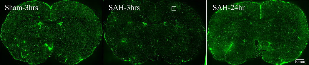
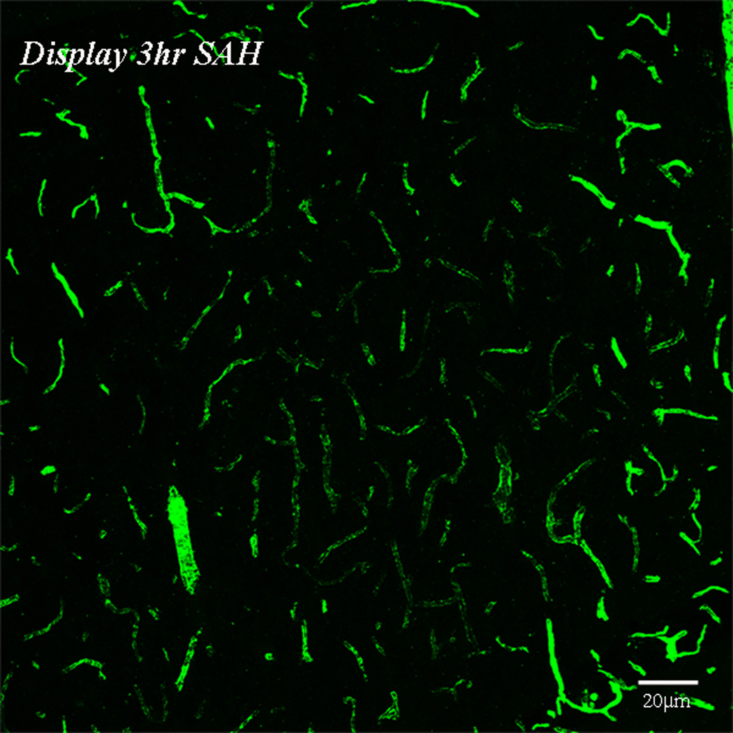
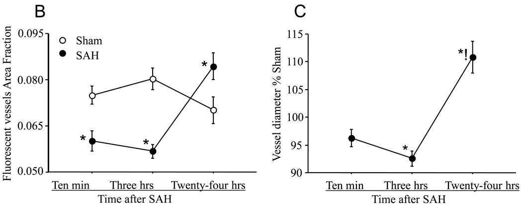
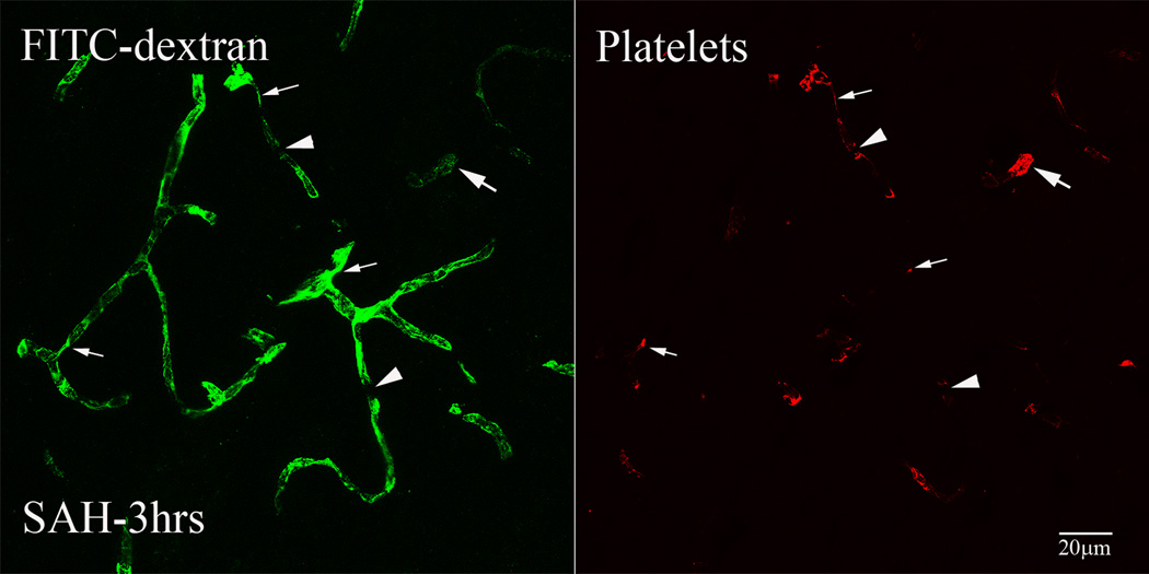
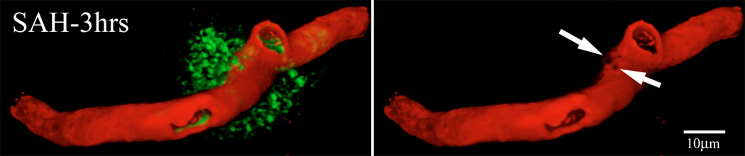
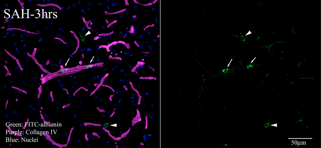
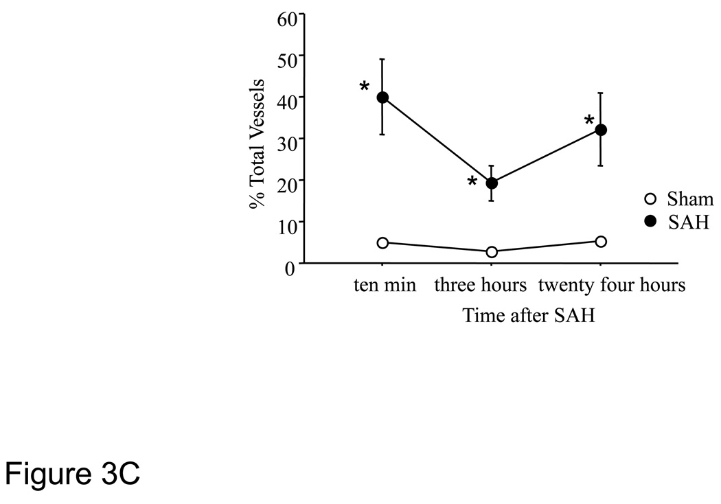
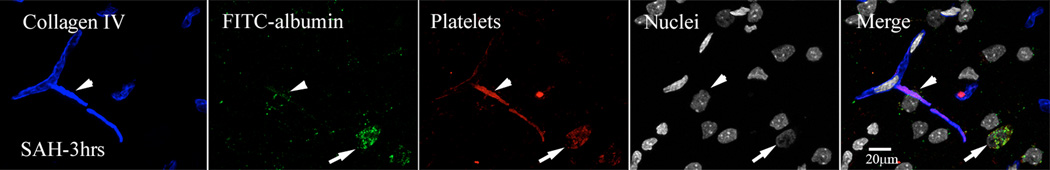
The pathophysiology of early ischemic injury after aneurysmal subarachnoid hemorrhage (SAH) is not understood. This study examined the acute effect of endovascular puncture-induced SAH on parenchymal vessel function in rat, using intravascular fluorescent tracers to assess flow and vascular permeability and immunostaining to assess structural integrity and to visualize platelet aggregates. In sham-operated animals, vessels were well filled with tracer administered 10s before sacrifice, and parenchymal escape of tracer was rare. At ten minutes and three hours after hemorrhage, patches of poor vascular filling were distributed throughout the forebrain. Close examination of these regions revealed short segments of narrowed diameter along many profiles. Most vascular profiles with reduced perfusion contained platelet aggregates and in addition showed focal loss of collagen IV, a principal component of basal lamina. In contrast, vessels were well filled at 24h post-hemorrhage, indicating that vascular perfusion had recovered. Parenchymal escape of intravascular tracer was detected at 10 min post-hemorrhage and later as plumes of fluorescence emanating into parenchyma from restricted microvascular foci. These data demonstrate that parenchymal microvessels are compromised in function by 10 min after SAH and identify focal microvascular constriction and local accumulation of luminal platelet aggregates as potential initiators of that compromise.


Figures

Figure-1
Figure-1. Perfusion deficits in parenchymal vessels after SAH
Animals were sacrificed at 10 minutes, 3 or 24 hours after Sham or SAH surgeries. Perfusion was visualized as the vascular presence of FITC-dextran injected 10 seconds before sacrifice. A: representative coronal brain sections from animals sacrificed at 3 hours after sham or 3 or 24 hours after SAH surgeries. Note the decrease in FITC-dextran labeled vessels at 3 hours after SAH and an increase at 24 hours as compared to sham. The boxed vessels in the in 3 hour SAH image are displayed in high magnification. B: Temporal change in area fraction of FITC-dextran positive vascular profiles during the first 24 hours after SAH. * significantly different than time matched sham cohorts at P<0.05. C: Temporal change in the diameter of FITC-dextran positive vascular profiles during the first 24 hours after SAH. Note small but significant increase in vessel diameter at 24 hour after SAH. Data are represented as % time matched shams. Mean ± sem, n=5 per time interval per surgery group. *: P<0.05.
Figure-2
Figure-2. Intraluminal platelet aggregates contribute in perfusion deficits after SAH
Brain sections from FITC-dextran-injected animals were immunostained for platelets. Shown is a representative 3D rendered stacked high magnification image from animal sacrifice 3 hours after SAH. Two types of vascular segments could be seen; vascular segments filled with platelet aggregates but contain little FITC-dextran label (arrows) and vascular segments with decreased diameter filled with platelet aggregates and FITC-dextran (thin arrows). Some FITC-dextran label vascular profiles appeared interrupted with platelet aggregates present at the interrupted edges (arrow heads).
Figure-3
Figure-3. Permeability changes in parenchymal vessels after SAH
Permeability was studied as extravasation of FITC-albumin injected 15 minutes before sacrifice. Brain sections were immunostained for collagen IV to delineate vessels. 3 D rendered stacked high magnification representative images from animals sacrificed 3 hours after SAH are shown. A: FITC-albumin leak from a collagen IV (red) immunostained vessel. B: A representative image showing green fluorescent vascular and parenchymal cells engaged in clean up process. Arrows: vascular green fluorescent cells, Arrow heads: parenchymal green fluorescent cells. C: FITC-albumin extravasation during the first 24 hours after SAH. Significantly greater number of vessels exhibited FITC-albumin extravasation at 10 minutes to 24 hours post-hemorrhage as compared to time matched sham cohorts. Data is representative as % time matched shams. Mean ± S.E.M, n=5 per time interval per surgery group. *: P<0.05.
Figure-4
Figure-4. Intraluminal platelet aggregates contribute in permeability changes after SAH
FITC-albumin injected brains were immunostained for platelets and collagen IV. Shown is a representative high magnification image from animal sacrifice 3 hours after SAH. Arrow heads: extravasation of FITC-Albumin and platelet (red) form a single site on collagen (blue) immunostained vessel. Arrows: Also note green fluorescent cells engaged in vascular and parenchymal clean up process.

References

    1. Akopov S, Sercombe R, Seylaz J. Cerebrovascular reactivity: role of endothelium/platelet/leukocyte interactions. Cerebrovasc Brain Metab Rev. 1996;8:11–94. - PubMed
    1. Bederson JB, Germano IM, Guarino L. Cortical blood flow and cerebral perfusion pressure in a new noncraniotomy model of subarachnoid hemorrhage in the rat. Stroke. 1995;26:1086–1091. - PubMed
    1. Bederson JB, Levy AL, Ding WH, Kahn R, DiPerna CA, Jenkins ALr, Vallabhajosyula P. Acute vasoconstriction after subarachnoid hemorrhage. Neurosurgery. 1998;42:352–360. - PubMed
    1. Doczi T. The pathogenetic and prognostic significance of blood-brain barrier damage at the acute stage of aneurysmal subarachnoid haemorrhage. Clinical and experimental studies. Acta Neurochir (Wien) 1985;77:110–132. - PubMed
    1. Doczi T, Joo F, Adam G, Bozoky B, Szerdahelyi P. Blood-brain barrier damage during the acute stage of subarachnoid hemorrhage, as exemplified by a new animal model. Neurosurgery. 1986;18:733–739. - PubMed

Publication types

Substances