

Review
doi: 10.1016/j.addr.2010.07.004.
Epub 2010 Jul 21.
Multifunctional agents for concurrent imaging and therapy in cardiovascular disease
Affiliations
- PMID: 20654664
- PMCID: PMC2974776
- DOI: 10.1016/j.addr.2010.07.004
Item in Clipboard
Review
Multifunctional agents for concurrent imaging and therapy in cardiovascular disease
Adv Drug Deliv Rev.
.
Abstract
The development of agents for the simultaneous detection and treatment of disease has recently gained significant attention. These multifunctional theranostic agents posses a number of advantages over their monofunctional counterparts, as they potentially allow for the concomitant determination of agent localization, release, and efficacy. Whereas the development of these agents for use in cancers has received the majority of the attention, their use in cardiovascular disease is steadily increasing. As such, this review summarized some of the most poignant recent advances in the development of theranostic agents for the treatment of this class of diseases.
Copyright © 2010 Elsevier B.V. All rights reserved.
Figures

MRI of abdominal aorta showing outline of segmented region of interest (ROI) (top), false-colored overlay of percent signal enhancement at time of treatment (middle), and 1 week post-treatment (bottom). The color overlays are thresholded at 10% enhancement to show some anatomic detail within the ROI. Reproduced with permission from [21].

Focal macrophage ablation. In vivo localization of the phototoxic nanoagent to carotid atheroma, as determined by intravital fluorescence microscopy. A) Fluorescence image in the AF750 channel demonstrating particle uptake by a carotid plaque. B) Fluorescence angiogram utilizing fluorescein-labeled dextran outlining the vasculature. C) Merged image of the two fluorescence channels.

Serial histological analysis of femoral arteries 2 weeks after balloon overstretch injury. Top, Treatment with ανβ3-integrin–targeted rapamycin nanoparticles. Bottom, Serial sections of injured femoral segment after exposure to ανβ3-integrin–targeted nanoparticles without drug. Serial cryosections stained with H&E represent 2-mm segments starting proximal to the injury (left) and ending distally (right). Lesion areas are depicted in green/orange and illustrate an irregular pattern of stenosis development and remodeling response along the injured vessel segments. Reproduced with permission from [35].

Localization of CREKA micelles in atherosclerotic plaques. (A) Serial cross-sections (5 μm thick) were stained with antibodies against CD31 (endothelial cells; Top), CD68 (macrophages and other lymphocytes; Middle), and fibrin(ogen) (Bottom). Representative microscopic fields are shown to illustrate the localization of micelle nanoparticles in the atherosclerotic plaque. Micelles are bound to the entire surface of the plaque with no apparent binding to the healthy portion of the vessel. CREKA targeted micelles also penetrate under the endothelial layer (CD31 staining) in the shoulder of the plaque (Inset) where there is high inflammation (CD68 staining) and the plaque is prone to rupture. Clotted plasma proteins are seen throughout the plaque and its surface [fibrin(ogen) staining]. (Left) Images were taken at a 10× magnification. (Scale bar, 200 μm.) (Right) Images were taken at a 150× magnification. (Scale bar, 20 μm.) (B) Fluorescence was not observed in the heart or lung, and only a small amount was seen in the kidney, spleen, and liver. Images were taken at a 20× magnification. (Scale bar, 100 μm.) Reproduced with permission from [37].
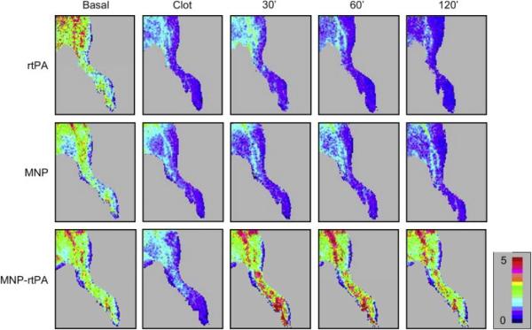
MNP-bound rtPA improved tissue perfusion in a rat embolic model. Hind limb skin tissue perfusion of the rat was measured by a laser Doppler perfusion imager. After clot lodging into the left iliac artery, rtPA (0.2 mg/kg; 0.27 U/kg), MNP-bound rtPA (0.2 mg/kg; 0.22 U/kg) or equivalent MNP (2.5 mg/kg) was administered from the right iliac arterial 5 min after introducing the clot. Reproduced with permission from [49].
Similar articles
-
Theranostic nanoparticles for cancer and cardiovascular applications.Pharm Res. 2014 Jun;31(6):1390-406. doi: 10.1007/s11095-013-1277-z. Epub 2014 Mar 5. Pharm Res. 2014. PMID: 24595494 Review.
-
Responsive theranostic systems: integration of diagnostic imaging agents and responsive controlled release drug delivery carriers.Acc Chem Res. 2011 Oct 18;44(10):1061-70. doi: 10.1021/ar2001777. Epub 2011 Sep 20. Acc Chem Res. 2011. PMID: 21932809 Free PMC article. Review.
-
Development and applications of photo-triggered theranostic agents.Adv Drug Deliv Rev. 2010 Aug 30;62(11):1094-124. doi: 10.1016/j.addr.2010.09.002. Epub 2010 Sep 19. Adv Drug Deliv Rev. 2010. PMID: 20858520 Free PMC article. Review.
-
Imaging and drug delivery using theranostic nanoparticles.Adv Drug Deliv Rev. 2010 Aug 30;62(11):1052-1063. doi: 10.1016/j.addr.2010.08.004. Epub 2010 Aug 13. Adv Drug Deliv Rev. 2010. PMID: 20709124 Free PMC article. Review.
-
Theranostic nanogels: multifunctional agents for simultaneous therapeutic delivery and diagnostic imaging.Nanoscale. 2024 Jul 25;16(29):14033-14056. doi: 10.1039/d4nr01423e. Nanoscale. 2024. PMID: 38990143 Review.
Cited by
-
Surface functionalized nanomaterial systems for targeted therapy of endocrine related tumors: a review of recent advancements.Drug Deliv. 2024 Dec;31(1):2390022. doi: 10.1080/10717544.2024.2390022. Epub 2024 Aug 13. Drug Deliv. 2024. PMID: 39138394 Free PMC article. Review.
-
Phase-shift, stimuli-responsive drug carriers for targeted delivery.Ther Deliv. 2011 Sep;2(9):1165-87. doi: 10.4155/tde.11.81. Ther Deliv. 2011. PMID: 22059114 Free PMC article. Review.
-
A non-invasive nanoparticles for multimodal imaging of ischemic myocardium in rats.J Nanobiotechnology. 2021 Mar 22;19(1):82. doi: 10.1186/s12951-021-00822-7. J Nanobiotechnology. 2021. PMID: 33752679 Free PMC article.
-
Near-infrared fluorescent probes in cancer imaging and therapy: an emerging field.Int J Nanomedicine. 2014 Mar 5;9:1347-65. doi: 10.2147/IJN.S60206. eCollection 2014. Int J Nanomedicine. 2014. PMID: 24648733 Free PMC article. Review.
-
Surface-modified nanotherapeutics targeting atherosclerosis.Biomater Sci. 2022 Sep 27;10(19):5459-5471. doi: 10.1039/d2bm00660j. Biomater Sci. 2022. PMID: 35980230 Free PMC article. Review.
References
-
- Kung HC, Hoyert DL, Xu J, Murphy SL. Deaths: final data for 2005. Natl Vital Stat Rep. 2008;56:1–120. - PubMed
-
- Jaffer FA, Nahrendorf M, Sosnovik D, Kelly KA, Aikawa E, Weissleder R. Cellular imaging of inflammation in atherosclerosis using magnetofluorescent nanomaterials. Mol Imaging. 2006;5:85–92. - PubMed
-
- Pande AN, Kohler RH, Aikawa E, Weissleder R, Jaffer FA. Detection of macrophage activity in atherosclerosis in vivo using multichannel, high-resolution laser scanning fluorescence microscopy. J Biomed Opt. 2006;11:021009. - PubMed
-
- Hyafil F, Cornily JC, Feig JE, Gordon R, Vucic E, Amirbekian V, Fisher EA, Fuster V, Feldman LJ, Fayad ZA. Noninvasive detection of macrophages using a nanoparticulate contrast agent for computed tomography. Nat Med. 2007;13:636–641. - PubMed
Publication types
MeSH terms
Substances
Grants and funding
LinkOut - more resources
Full Text Sources
Medical
Research Materials
