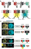
Fluorescent protein complementation assays: new tools to study G protein-coupled receptor oligomerization and GPCR-mediated signaling
- PMID: 20654687
- PMCID: PMC2990800
- DOI: 10.1016/j.mce.2010.07.011
Fluorescent protein complementation assays: new tools to study G protein-coupled receptor oligomerization and GPCR-mediated signaling
Abstract
G protein-coupled receptor (GPCR) signaling is mediated by protein-protein interactions at multiple levels. The characterization of the corresponding protein complexes is therefore paramount to the basic understanding of GPCR-mediated signal transduction. The number of documented interactions involving GPCRs is rapidly growing, and appreciating the functional significance of these complexes is clearly the next challenge. New experimental approaches including protein complementation assays (PCAs) have recently been used to examine the composition, plasma membrane targeting, and desensitization of protein complexes involved in GPCR signaling. These methods also hold promise for better understanding of drug-induced effects on GPCR interactions. This review focuses on the application of fluorescent PCAs for the study of GPCR signaling. Potential applications of PCAs in high-content screens are also presented. Non-fluorescent PCA techniques as well as combined assays for the detection of ternary and quaternary protein complexes are briefly discussed.
Copyright © 2010 Elsevier Ireland Ltd. All rights reserved.
Figures


Similar articles
-
Fluorescent and bioluminescent protein-fragment complementation assays in the study of G protein-coupled receptor oligomerization and signaling.Mol Pharmacol. 2009 Apr;75(4):733-9. doi: 10.1124/mol.108.053819. Epub 2009 Jan 13. Mol Pharmacol. 2009. PMID: 19141658 Free PMC article. Review.
-
Luminescence- and Fluorescence-Based Complementation Assays to Screen for GPCR Oligomerization: Current State of the Art.Int J Mol Sci. 2019 Jun 17;20(12):2958. doi: 10.3390/ijms20122958. Int J Mol Sci. 2019. PMID: 31213021 Free PMC article. Review.
-
On the expanding terminology in the GPCR field: the meaning of receptor mosaics and receptor heteromers.J Recept Signal Transduct Res. 2010 Oct;30(5):287-303. doi: 10.3109/10799891003786226. J Recept Signal Transduct Res. 2010. PMID: 20429829 Free PMC article. Review.
-
The Dynamics of GPCR Oligomerization and Their Functional Consequences.Int Rev Cell Mol Biol. 2018;338:141-171. doi: 10.1016/bs.ircmb.2018.02.005. Epub 2018 Apr 7. Int Rev Cell Mol Biol. 2018. PMID: 29699691 Review.
-
Oligomerization of GPCRs involved in endocrine regulation.J Mol Endocrinol. 2016 Jul;57(1):R59-80. doi: 10.1530/JME-16-0049. Epub 2016 May 5. J Mol Endocrinol. 2016. PMID: 27151573 Review.
Cited by
-
Methods used to study the oligomeric structure of G-protein-coupled receptors.Biosci Rep. 2017 Apr 20;37(2):BSR20160547. doi: 10.1042/BSR20160547. Print 2017 Apr 30. Biosci Rep. 2017. PMID: 28062602 Free PMC article. Review.
-
Receptor oligomerization in family B1 of G-protein-coupled receptors: focus on BRET investigations and the link between GPCR oligomerization and binding cooperativity.Front Endocrinol (Lausanne). 2012 May 7;3:62. doi: 10.3389/fendo.2012.00062. eCollection 2012. Front Endocrinol (Lausanne). 2012. PMID: 22649424 Free PMC article.
-
Bioluminescence resonance energy transfer methods to study G protein-coupled receptor-receptor tyrosine kinase heteroreceptor complexes.Methods Cell Biol. 2013;117:141-64. doi: 10.1016/B978-0-12-408143-7.00008-6. Methods Cell Biol. 2013. PMID: 24143976 Free PMC article.
-
Principles and Design of Molecular Tools for Sensing and Perturbing Cell Surface Receptor Activity.Chem Rev. 2025 Mar 12;125(5):2665-2702. doi: 10.1021/acs.chemrev.4c00582. Epub 2025 Feb 25. Chem Rev. 2025. PMID: 39999110 Review.
-
Oligomerization of Family B GPCRs: Exploration in Inter-Family Oligomer Formation.Front Endocrinol (Lausanne). 2015 Feb 2;6:10. doi: 10.3389/fendo.2015.00010. eCollection 2015. Front Endocrinol (Lausanne). 2015. PMID: 25699019 Free PMC article. Review.
References
-
- Antonelli T, Fuxe K, Agnati L, Mazzoni E, Tanganelli S, Tomasini MC, Ferraro L. Experimental studies and theoretical aspects on A2A/D2 receptor interactions in a model of Parkinson’s disease. Relevance for L-dopa induced dyskinesias. J Neurol Sci. 2006;248:16–22. - PubMed
-
- Armstrong D, Strange PG. Dopamine D2 receptor dimer formation: evidence from ligand binding. J Biol Chem. 2001;276:22621–9. - PubMed
-
- Baragli A, Grieco ML, Trieu P, Villeneuve LR, Hebert TE. Heterodimers of adenylyl cyclases 2 and 5 show enhanced functional responses in the presence of Galpha s. Cell Signal. 2008;20:480–92. - PubMed
-
- Bulenger S, Marullo S, Bouvier M. Emerging role of homo- and heterodimerization in G-protein-coupled receptor biosynthesis and maturation. Trends Pharmacol Sci. 2005;26:131–7. - PubMed
Publication types
MeSH terms
Substances
Grants and funding
LinkOut - more resources
Full Text Sources

