Review
. 2010 Oct;1804(10):1943-53.
doi: 10.1016/j.bbapap.2010.07.016. Epub 2010 Jul 21.

Mechanistic similarity and diversity among the guanidine-modifying members of the pentein superfamily

Affiliations
Review

Mechanistic similarity and diversity among the guanidine-modifying members of the pentein superfamily

Thomas Linsky et al. Biochim Biophys Acta. 2010 Oct.

Abstract

The pentein superfamily is a mechanistically diverse superfamily encompassing both noncatalytic proteins and enzymes that catalyze hydrolase, dihydrolase and amidinotransfer reactions on guanidine substrates. Despite generally low sequence identity, they possess a conserved structural fold and display common mechanistic themes in catalysis. The structurally characterized catalytic penteins possess a conserved core of residues that include a Cys, His and two polar, guanidine-binding residues. All known catalytic penteins use the core Cys to attack the substrate's guanidine moiety to form a covalent thiouronium adduct and all cleave one or more of the guanidine C--N bonds. The mechanistic information compiled to date supports the hypothesis that this superfamily may have evolved divergently from a catalytically promiscuous ancestor.


Figures

Fig. 1
Fig. 1
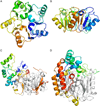
The pentein fold consists of five αββαβ subdomains symmetrically arranged around afivefold axis of pseudosymmetry. A) Top-down viewofribosome anti-association factor eIF6 (PDB ID: 1G61). B) Side view of eIF6. C) Side view of arginine deiminase (PDB ID: 2A9G). D) Side view of succinylarginine dihydrolase (PDB ID: 1YNI). E) Side view of arginine: glycine amidinotransferase (PDB ID: 4JDW). F) Side view of PAD4 (PDB ID: 1WDA). In C–F, the conserved pentein core is colored gray, while additional inserts and loops are colored by amino acid residue number.
Fig. 1
Fig. 1
The pentein fold consists of five αββαβ subdomains symmetrically arranged around afivefold axis of pseudosymmetry. A) Top-down viewofribosome anti-association factor eIF6 (PDB ID: 1G61). B) Side view of eIF6. C) Side view of arginine deiminase (PDB ID: 2A9G). D) Side view of succinylarginine dihydrolase (PDB ID: 1YNI). E) Side view of arginine: glycine amidinotransferase (PDB ID: 4JDW). F) Side view of PAD4 (PDB ID: 1WDA). In C–F, the conserved pentein core is colored gray, while additional inserts and loops are colored by amino acid residue number.
Fig. 2
Fig. 2
Reactions catalyzed by penteins. The guanidine substrate binds and reacts with the core Cys residue to form a thiouronium intermediate, which is used in the hydrolase, amidinotransferase and dihydrolase reactions. It is assumed in this figure that the active-site Cys is deprotonated prior to formation of the alkylthiouronium adduct. However, the protonation model at work is unclear (see Fig. 6).
Fig. 3
Fig. 3
Generic active-site diagram. Residues not conserved are shown in gray. R1=Nδ of arginine substrate in hydrolases, R2=Nδ of arginine substrate in amidinotransferases.
Fig. 4
Fig. 4
Active-site diagram showing core catalytic residues for A) ADI, AgD and PAD, B) AGAT and AIAT, C) DDAH and D) SADH. Note that the terms “lateral” and “anterior” refer to the position of the residue relative to the substrate arginine’s Nδ—Cζ bond in the hydrolases and dihydrolases.
Fig. 5
Fig. 5
Proposed reaction mechanisms for A) hydrolases, B) amidinotransferases, C) dihydrolases.
Fig. 6
Fig. 6
Proposed protonation states of pentein core Cys.
Fig. 7
Fig. 7
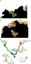
Structures of A) hydrolase (Green) and B) amidinotransferase (Orange) active sites, and C) an overlay of the two active sites. The hydrolase is ADI from P. aeruginosa (PDB ID: 2a9g) and the amidinotransferase is human AGAT (PDBID: 4jdw). Substrate is positioned at different relative angles, presumable to facilitate cleavage of a different C–N bond. In both structures, the core Cys is mutated to Ala to enable formation of a stable substrate complex.

Similar articles

Cited by

References

    1. Groft CM, Beckmann R, Sali A, Burley SK. Crystal structures of ribosome anti-association factor IF6. Nature Structural & Molecular Biology. 2000 Dec;7:1156–1164. - PubMed
    1. Paoli M. An elusive propeller-like fold. Nature Structural Biology. 2001 Sep;8:744–745. - PubMed
    1. Teichmann SA, Murzin AG, Chothia C, Determination of protein function. evolution and interactions by structural genomics. Current Opinion in Structural Biology. 2001 Jun;11:354–363. - PubMed
    1. Hartzoulakis B, Rossiter S, Gill H, O’Hara B, Steinke E, Gane PJ, Hurtado-Guerrero R, Leiper JM, Vallance P, Rust JM, Selwood DL. Discovery of inhibitors of the pentein superfamily protein dimethylarginine dimethylaminohydrolase (DDAH) by virtual screening and hit analysis. Bioorganic & Medicinal Chemistry Letters. 2007 Jul;17:3953–3956. - PubMed
    1. Groft CM, Beckmann R, Sali A, Burley SK. Crystal structures of ribosome anti-association factor IF6. Nature Structural Biology. 2000 Dec;7:1156–1164. - PubMed

Publication types

LinkOut - more resources