

Association between amygdala response to emotional faces and social anxiety in autism spectrum disorders
- PMID: 20655320
- PMCID: PMC3426451
- DOI: 10.1016/j.neuropsychologia.2010.07.022
Association between amygdala response to emotional faces and social anxiety in autism spectrum disorders
Abstract
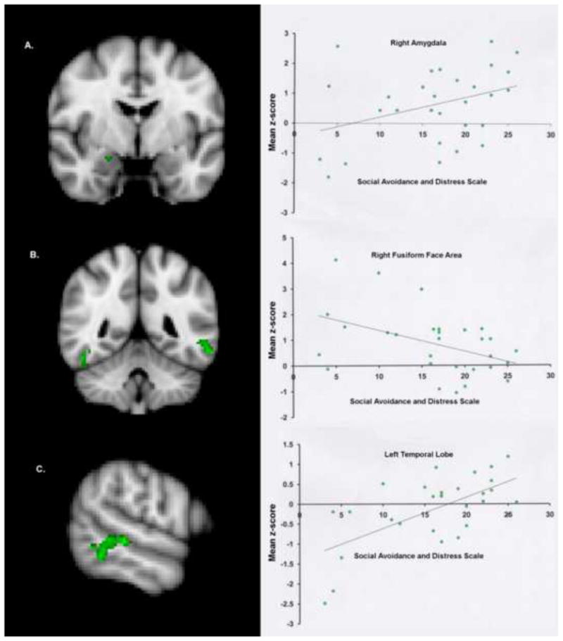
Difficulty interpreting facial expressions has been reported in autism spectrum disorders (ASD) and is thought to be associated with amygdala abnormalities. To further explore the neural basis of abnormal emotional face processing in ASD, we conducted an fMRI study of emotional face matching in high-functioning adults with ASD and age, IQ, and gender matched controls. In addition, we investigated whether there was a relationship between self-reported social anxiety and fMRI activation. During fMRI scanning, study participants were instructed to match facial expressions depicting fear or anger. The control condition was a comparable shape-matching task. The control group evidenced significantly increased left prefrontal activation and decreased activation in the occipital lobes compared to the ASD group during emotional face matching. Further, within the ASD group, greater social anxiety was associated with increased activation in right amygdala and left middle temporal gyrus, and decreased activation in the fusiform face area. These results indicate that level of social anxiety mediates the neural response to emotional face perception in ASD.
Copyright © 2010 Elsevier Ltd. All rights reserved.
Figures

Similar articles
-
Neural circuitry of emotional face processing in autism spectrum disorders.J Psychiatry Neurosci. 2010 Mar;35(2):105-14. doi: 10.1503/jpn.090085. J Psychiatry Neurosci. 2010. PMID: 20184808 Free PMC article.
-
fMRI evidence of neural abnormalities in the subcortical face processing system in ASD.Neuroimage. 2011 Jan 1;54(1):697-704. doi: 10.1016/j.neuroimage.2010.07.037. Epub 2010 Jul 23. Neuroimage. 2011. PMID: 20656041 Free PMC article.
-
Effects of intranasal oxytocin on the neural basis of face processing in autism spectrum disorder.Biol Psychiatry. 2013 Aug 1;74(3):164-71. doi: 10.1016/j.biopsych.2013.02.007. Epub 2013 Mar 16. Biol Psychiatry. 2013. PMID: 23510581 Clinical Trial.
-
Research review: Constraining heterogeneity: the social brain and its development in autism spectrum disorder.J Child Psychol Psychiatry. 2011 Jun;52(6):631-44. doi: 10.1111/j.1469-7610.2010.02349.x. Epub 2011 Jan 19. J Child Psychol Psychiatry. 2011. PMID: 21244421 Free PMC article. Review.
-
Social cognition in schizophrenia: a review of face processing.Br Med Bull. 2008;88(1):43-58. doi: 10.1093/bmb/ldn035. Epub 2008 Sep 23. Br Med Bull. 2008. PMID: 18812413 Review.
Cited by
-
Autistic and non-autistic individuals show the same amygdala activity during emotional face processing.Mol Autism. 2024 Jan 10;15(1):2. doi: 10.1186/s13229-024-00582-9. Mol Autism. 2024. PMID: 38200601 Free PMC article.
-
Fear of Negative Evaluation Influences Eye Gaze in Adolescents with Autism Spectrum Disorder: A Pilot Study.J Autism Dev Disord. 2015 Nov;45(11):3446-57. doi: 10.1007/s10803-014-2349-6. J Autism Dev Disord. 2015. PMID: 25578337
-
RDoC-based categorization of amygdala functions and its implications in autism.Neurosci Biobehav Rev. 2018 Jul;90:115-129. doi: 10.1016/j.neubiorev.2018.04.007. Epub 2018 Apr 13. Neurosci Biobehav Rev. 2018. PMID: 29660417 Free PMC article. Review.
-
Functional connectivity within an anxiety network and associations with anxiety symptom severity in middle-aged adults with and without autism.Autism Res. 2021 Oct;14(10):2100-2112. doi: 10.1002/aur.2579. Epub 2021 Jul 15. Autism Res. 2021. PMID: 34264028 Free PMC article.
-
Age-related reduction in trait anxiety: Behavioral and neural evidence of automaticity in negative facial emotion processing.Neuroimage. 2023 Aug 1;276:120207. doi: 10.1016/j.neuroimage.2023.120207. Epub 2023 May 30. Neuroimage. 2023. PMID: 37263454 Free PMC article.
References
-
- Ashwin C, Baron-Cohen S, Wheelwright S, O’Riordan M, Bullmore ET. Differential activation of the amygdala and the ‘social brain’ during fearful face-processing in Asperger Syndrome. Neuropsychologia 2006 - PubMed
-
- Ashwin C, Chapman E, Colle L, Baron-Cohen S. Impaired recognition of negative basic emotions in autism: a test of the amygdala theory. Soc Neurosci. 2006;1(3–4):349–363. - PubMed
-
- Baron-Cohen S, Ring HA, Bullmore ET, Wheelwright S, Ashwin C, Williams SC. The amygdala theory of autism. Neurosci Biobehav Rev. 2000;24(3):355–364. - PubMed
-
- Belmonte MK, Yurgelun-Todd DA. Functional anatomy of impaired selective attention and compensatory processing in autism. Brain Res Cogn Brain Res. 2003;17(3):651–664. - PubMed
Publication types
MeSH terms
Substances
Grants and funding
LinkOut - more resources
Full Text Sources
Other Literature Sources
Medical
