Review
. 2010 Sep;31(9):418-26.
doi: 10.1016/j.tips.2010.06.004. Epub 2010 Jul 23.

Structural perspectives on secondary active transporters

Affiliations
Review

Structural perspectives on secondary active transporters

Olga Boudker et al. Trends Pharmacol Sci. 2010 Sep.

Abstract

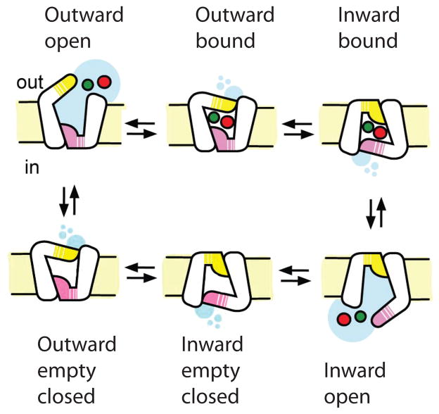
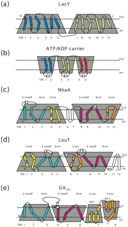
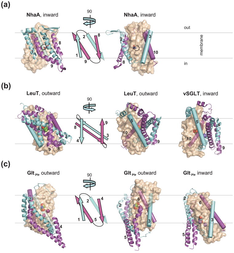
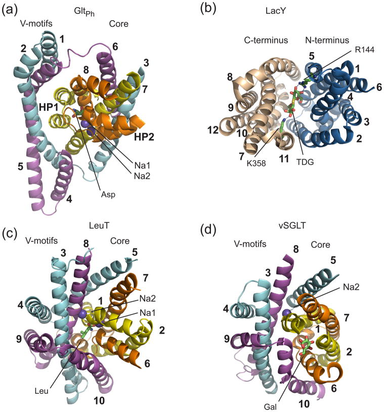
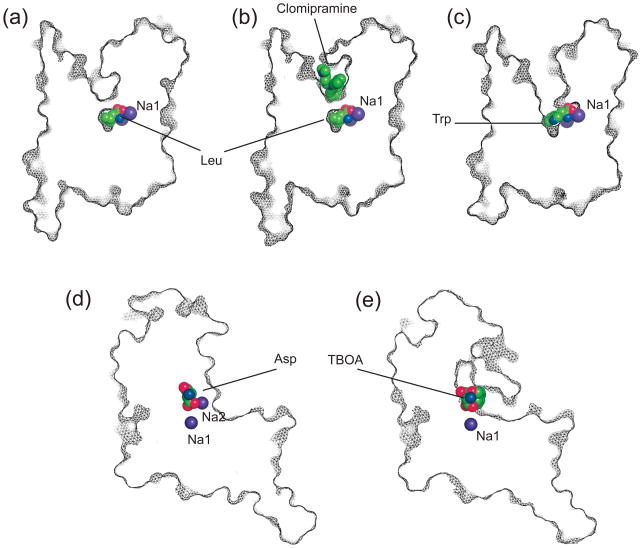
Secondary active transporters catalyze the concentrative transport of substrates across lipid membranes by harnessing the energy of electrochemical ion gradients. These transporters bind their ligands on one side of the membrane, and undergo a global conformational change to release them on the other side of the membrane. Over the last few years, crystal structures have captured several bacterial secondary transporters in different states along their transport cycle, providing insight into possible molecular mechanisms. In this review, we summarize recent findings focusing on the emerging structural and mechanistic similarities between evolutionary diverse transporters. We also discuss the structural basis of substrate binding, ion coupling and inhibition viewed from the perspective of these similarities.


Figures

Figure 1
Figure 1. Alternating access mechanism for an ion/substrate symport
The cycle starts with an empty outward facing transporter, which binds the substrate and coupled ion (red and green spheres, respectively) from the extracellular solution in a reaction coupled to the closure of the gate (yellow). The transporter undergoes a conformational transition into an inward facing state in which the extracellular gate can no longer open while opening of the intracellular gate (pink) is enabled. The release of the substrate and ion into the cytoplasm yields an empty inward facing state, and isomerization back into the outward facing state completes the cycle.
Figure 2
Figure 2. Membrane topology of transporters with parallel and inverted structural repeats
The topologies are shown for the MFS transporter LacY (a), ATP/ADP carrier (b), NhaA (c), LeuT (d) and GltPh (e). The structural repeats are highlighted by the shaded trapezoids, which emphasize their relative orientation. The N- and C-terminal TM segments comprising the transporter cores in GltPh, NhaA and LeuT are colored yellow and orange, respectively. The remaining N- and C-terminal portions of the repeats are colored cyan and magenta, respectively. The segments that are not part of the symmetrical repeats are white. Non-helical regions in the middle of the core TMs are shown as lines.
Figure 3
Figure 3. Architecture of the transporters with inverted structural repeats
(a) NhaA (PDB code 1zcd); (b) LeuT in the outward-facing state (PDB code 2a65) and vSGLT in the inward-facing state (PDB code 3dh4); (c) GltPh in the outward- (PDB code 2nwx) and inward- (PDB code 3kbc) facing states. The leftmost panels show a single protomer viewed in the membrane plane from the side of the V-motifs with cartoon topologies of the V-motifs shown immediately to the right. In the right panels the structures are rotated by about 90 °, as indicated by the arrows. The V-motifs and the arms TM-s are colored as in Figure 2 and shown in cartoon representation and as cylinders, respectively. The cores are shown in transparent surface representation and colored wheat; the bound substrates and ions are shown as spheres. In NhaA, D164 is shown as sticks and black circles indicate expected site of ion binding. This and other figures were prepared in PyMol [84].
Figure 4
Figure 4. Binding sites
(a) GltPh; (b) LacY (PDB code 1pv7); (c) LeuT; (d) vSGLT. Transporters are shown in cartoon representation and colored as in Figure 2. LeuT and vSGLT are viewed from the extracellular space and LacY is viewed from the cytoplasm; GltPh is tilted to reveal the binding site. The TMs that are not part of the symmetrical repeats, extracellular loops, and parts of TM helices are removed for clarity. The substrates and two coordinating residues in LacY are shown stick representation and bound Na+ ions are shown as purple balls.
Figure 5
Figure 5. Competitive blockers and non-competitive inhibitors
(a) LeuT bound to leucine; (b) LeuT bound to leucine and non-competitive inhibitor clomipramine (PDB code 2qei); (c) LeuT bound to competitive blocker tryptophan (PDB code 3f3a); (d) GltPh bound to aspartate; (e) GltPh bound to competitive blocker TBOA (PDB code 2nww). Shown are the thin slices through the transporter protomers in surface representation and viewed in the membrane plain. Bound ligands are emphasized as balls.

References

    1. Saier MH, Jr, et al. TCDB: the Transporter Classification Database for membrane transport protein analyses and information. Nucleic Acids Res. 2006;34:D181–186. - PMC - PubMed
    1. Hediger MA, et al. The ABCs of solute carriers: physiological, pathological and therapeutic implications of human membrane transport proteinsIntroduction. Pflugers Arch. 2004;447:465–468. - PubMed
    1. Lolkema JS, Slotboom DJ. Classification of 29 families of secondary transport proteins into a single structural class using hydropathy profile analysis. J Mol Biol. 2003;327:901–909. - PubMed
    1. Brett CL, et al. Evolutionary origins of eukaryotic sodium/proton exchangers. Am J Physiol Cell Physiol. 2005;288:C223–239. - PubMed
    1. Lolkema JS, Slotboom DJ. The major amino acid transporter superfamily has a similar core structure as Na+-galactose and Na+-leucine transporters. Mol Membr Biol. 2008;25:567–570. - PubMed

MeSH terms