. 2010 Jul 7;99(1):115-23.
doi: 10.1016/j.bpj.2010.04.011.

Actomyosin tension exerted on the nucleus through nesprin-1 connections influences endothelial cell adhesion, migration, and cyclic strain-induced reorientation

Affiliations

Actomyosin tension exerted on the nucleus through nesprin-1 connections influences endothelial cell adhesion, migration, and cyclic strain-induced reorientation

T J Chancellor et al. Biophys J. .

Erratum in

  • Biophys J. 2010 Aug 9;99(4):1329

Abstract

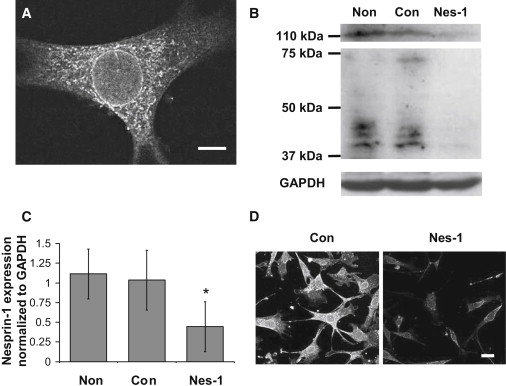
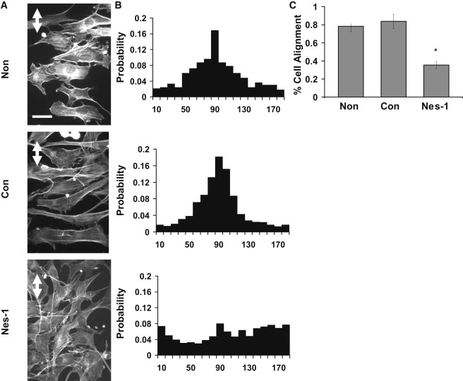
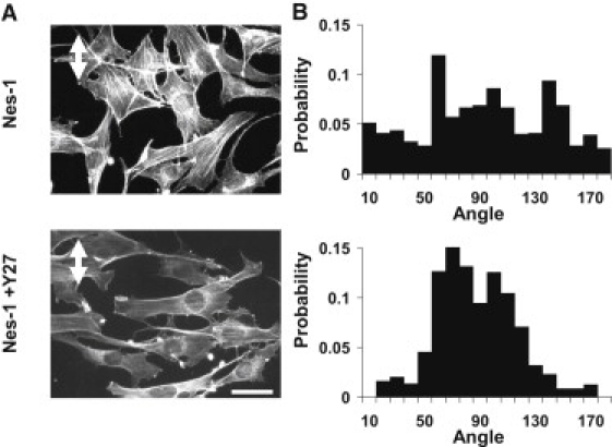
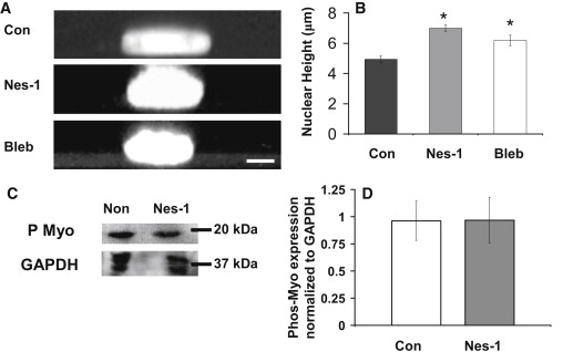
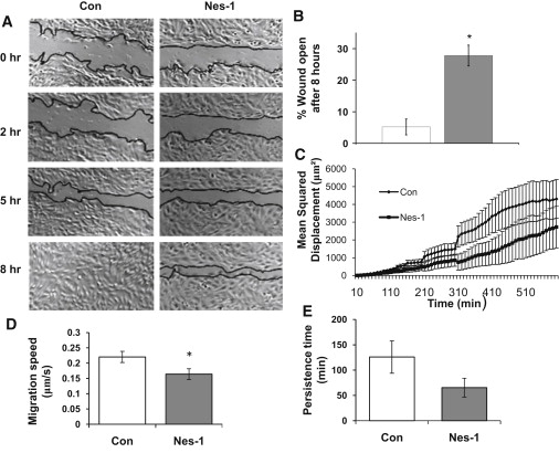
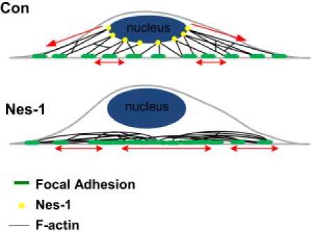
Endothelial cell polarization and directional migration is required for angiogenesis. Polarization and motility requires not only local cytoskeletal remodeling but also the motion of intracellular organelles such as the nucleus. However, the physiological significance of nuclear positioning in the endothelial cell has remained largely unexplored. Here, we show that siRNA knockdown of nesprin-1, a protein present in the linker of nucleus to cytoskeleton complex, abolished the reorientation of endothelial cells in response to cyclic strain. Confocal imaging revealed that the nuclear height is substantially increased in nesprin-1 depleted cells, similar to myosin inhibited cells. Nesprin-1 depletion increased the number of focal adhesions and substrate traction while decreasing the speed of cell migration; however, there was no detectable change in nonmuscle myosin II activity in nesprin-1 deficient cells. Together, these results are consistent with a model in which the nucleus balances a portion of the actomyosin tension in the cell. In the absence of nesprin-1, actomyosin tension is balanced by the substrate, leading to abnormal adhesion, migration, and cyclic strain-induced reorientation.


Figures

Figure 1
Figure 1
Transfection of HUVECs with siRNA targeting nesprin-1 results in a significant reduction in nesprin-1 expression. (A) Confocal fluorescence image of HUVEC immunostained for nesprin-1. Nesprin-1 localizes to the nuclear membrane. Scale bar = 5 μm. (B) Western blot analysis of nesprin-1 expression in HUVECs. HUVECs transfected with siRNA targeting nesprin-1 (Nes-1) show a significant reduction in nesprin-1 expression as compared to nontransfected cells (Non) and cells transfected with control siRNA (Con). (C) Quantification of nesprin-1 expression relative to GAPDH expression demonstrating a significant decrease in siRNA transfected cells. Error bars represent SE from three different experiments. p < .05 (D) Low magnification confocal fluorescence images of HUVECs immunostained for nesprin-1. A significant reduction in nesprin-1 expression is observed in cells transfected with nesprin-1 targeting siRNA (images were acquired at the same laser power, photomultiplier gain, and magnification). Scale bar = 25 μm.
Figure 2
Figure 2
Nesprin-1 deficient HUVECs are unable to align in response to uniaxial cyclic strain. (A) HUVECs cultured on flexible silicon membranes coated with fibronectin were exposed to 10% cyclic, uniaxial strain at 0.5 Hz, fixed and stained with Alexa-phalloidin to visualize F-actin stress fibers. Nontransfected HUVECs and cells expressing control siRNA oriented perpendicular to the strain direction (strain direction is marked by white double arrow) whereas cells transfected with nesprin-1 targeting siRNA did not align in any preferred direction. Stress fibers were observed predominantly perpendicular to the strain direction except in nesprin-1 deficient cells. Scale bar = 50 μm. (B) Probability distributions of cell angle measured relative to strain axis. A clear preference for a direction perpendicular to the strain axis is observed in the distribution for nontransfected and control siRNA transfected cells; the distribution is random for nesprin-1 siRNA transfected cells. The distribution was quantified from pooled data from three independent experiments corresponding to 300 cells per condition. (C) Quantification of the reorientation response. The data is presented as percentage of cells that reoriented 90° ± 30° relative to the strain direction similar to the approach in Ghosh et al. (33). Error bars represent SE from three different experiments. p < 0.01.
Figure 3
Figure 3
Nesprin-1 depletion results in increased cell traction and focal adhesions. (A) The area per FA was unchanged between cells transfected with control and nesprin-1 targeting siRNA, whereas the number of FAs (B) increased in nesprin-1 deficient cells (p < 0.05). (C) Representative phase contrast images and traction stress maps of cells transfected with nesprin-1 targeting and control siRNA. Scale bar = 200 μm. (D) Surface strain energy is increased in nesprin-1 deficient cells compared to control cells (p < 0.05). Error bars represent SE from three different experiments. p < 0.01. A minimum of eight cells were measured for each condition.
Figure 4
Figure 4
Rho kinase inhibition restores the reorientation response in nesprin-1 deficient cells. (A) After 18 h of 10% cyclic, uniaxial strain at 0.5 Hz (strain direction is marked by white double arrow), cells transfected with nesprin-1 targeting siRNA did not align in any preferred direction. Pretreatment of cells with Y27632 for 30 min followed by washout and cyclic stretching restored the reorientation response of nesprin-1 deficient cells. Scale bar = 50 μm. (B) Probability distributions of cell angle measured relative to strain axis. Y27632-treated nesprin-1 deficient cells have a much higher probability of reorienting perpendicular to the applied strain. The distribution was quantified from pooled data from two independent experiments corresponding to 150 cells per condition.
Figure 5
Figure 5
Nuclear height increases in nesprin-1 deficient HUVECs. (A) Representative Z-stack images generated with confocal microscopy show an increase in nuclear height in nesprin-1 deficient and blebbistatin (bleb)-treated cells. Hoechst33342 was used to stain the nucleus. Scale bar = 5 μm. (B) Plot quantifies the increase in nuclear height of nesprin-1 deficient HUVECs and HUVECs treated with blebbistatin. Error bars represent SE; p < 0.05 (each statistical comparison is with Con). A minimum of 15 cells were measured for each condition. (C) Western blot of phosphorylated myosin (P Myo) in nontransfected and siRNA transfected cells. (D) Quantification shows no difference in myosin II light chain phosphorylation. Error bars represent SE from three different experiments.
Figure 6
Figure 6
Nesprin-1 deficient HUVECs have decreased wound healing rates, single-cell speed, and persistence times. (A) Phase contrast images of HUVECs at 0, 2, 5, and 8 h after wounding are shown. Wound edges are marked in black. Scale bar = 200 μm. (B) Plot shows the unhealed percentage of the original wound for HUVECs transfected with control and nesprin-1 siRNA at 8 h. Error bars represent SE from three different experiments. p < 0.01. (C) Plot shows MSD calculated using single-cell trajectories and pooled together from at least 10 different cells. Error bars represent SE. MSD is decreased significantly in nesprin-1 deficient cells. (D) Individual cell migration speed, and (E) persistence time is decreased in nesprin-1 deficient cells; the decrease in persistence time is not statistically significant. At least 17 cells were analyzed for each condition in the motility experiments. p < 0.05.
Figure 7
Figure 7
Physical model for nuclear-actomyosin force balance. In control cells (top) the actomyosin tension is balanced in part by the nucleus due to mechanical links mediated by nesprin-1. In the absence of nesprin-1 (bottom), the forces are balanced by the substrate at an increased number of focal adhesions even though myosin II activity is unchanged.

References

    1. Huang S., Ingber D.E. The structural and mechanical complexity of cell-growth control. Nat. Cell Biol. 1999;1:E131–E138. - PubMed
    1. Lauffenburger D.A., Horwitz A.F. Cell migration: a physically integrated molecular process. Cell. 1996;84:359–369. - PubMed
    1. Ferrara N., Gerber H.P., LeCouter J. The biology of VEGF and its receptors. Nat. Med. 2003;9:669–676. - PubMed
    1. Starr D.A. A nuclear-envelope bridge positions nuclei and moves chromosomes. J. Cell Sci. 2009;122:577–586. - PMC - PubMed
    1. Hale C.M., Shrestha A.L., Wirtz D. Dysfunctional connections between the nucleus and the actin and microtubule networks in laminopathic models. Biophys. J. 2008;95:5462–5475. - PMC - PubMed

Publication types

MeSH terms