

Thermodynamics of polyethylenimine-DNA binding and DNA condensation
- PMID: 20655848
- PMCID: PMC2895367
- DOI: 10.1016/j.bpj.2010.04.016
Thermodynamics of polyethylenimine-DNA binding and DNA condensation
Abstract
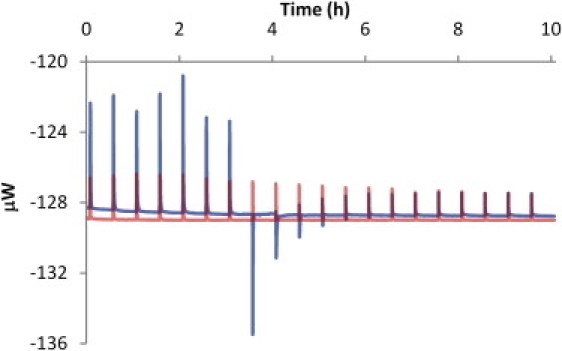
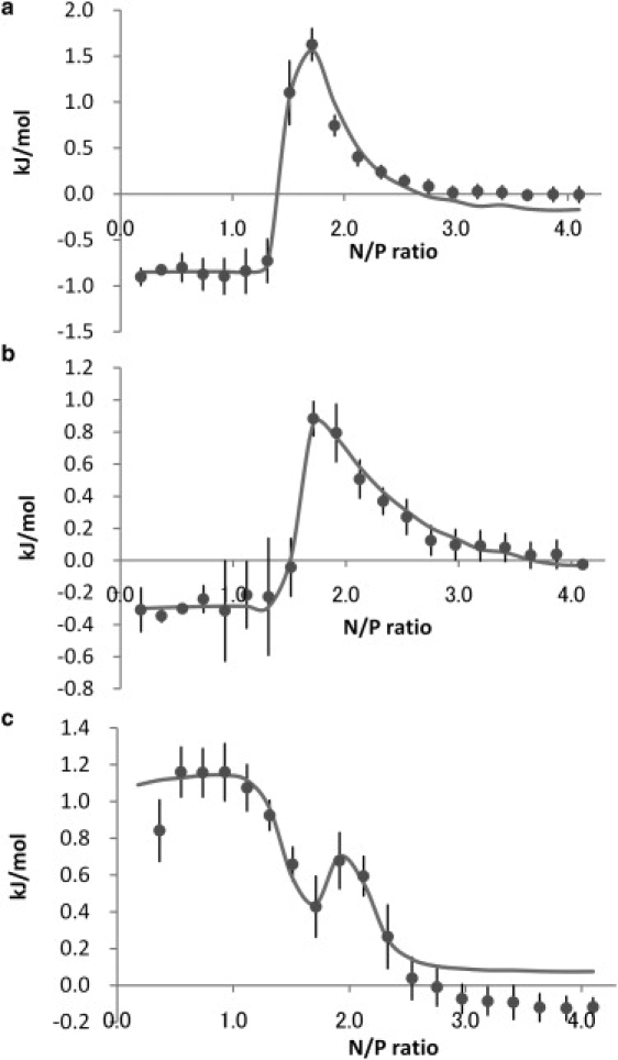
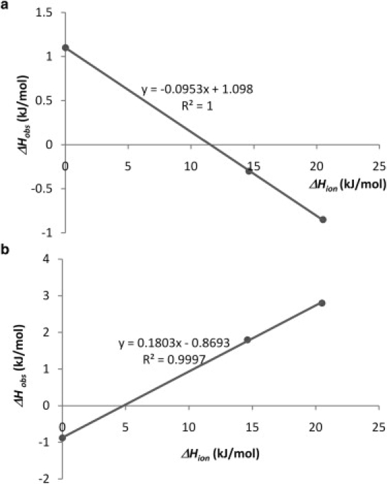
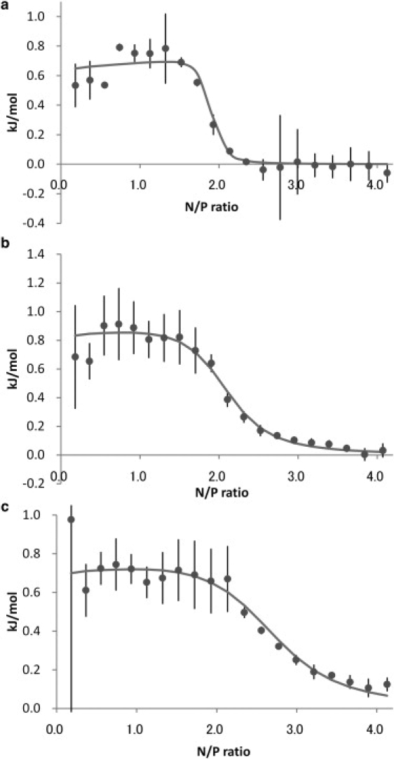
In this study, polyethylenimine (PEI) binding to DNA was examined by isothermal titration calorimetry. Two types of binding modes were found to describe the interactions between these polyelectrolytes in buffers and in water. One type of binding involves PEI binding to the DNA groove because the enthalpy change of this binding mode is positive, and PEI is deprotonated to bind to DNA. Another likely binding mode involves external binding of PEI to the DNA phosphate backbone, accompanied with DNA condensation. The enthalpy change is negative and PEI is protonated when it binds to DNA in this mode. The intrinsic enthalpy change of first binding mode is 1.1 kJ/mol and -0.88 kJ/mol for the second binding mode. This result implies that the PEI is rearranged from the groove to the phosphate backbone of DNA when DNA is condensed. The mechanism of DNA condensation caused by PEI is discussed in this study.
Copyright 2010 Biophysical Society. Published by Elsevier Inc. All rights reserved.
Figures
References
-
- Godbey W.T., Wu K.K., Mikos A.G. Poly(ethylenimine) and its role in gene delivery. J. Control. Release. 1999;60:149–160. - PubMed
-
- Godbey W.T., Wu K.K., Mikos A.G. Improved packing of poly(ethylenimine)/DNA complexes increases transfection efficiency. Gene Ther. 1999;6:1380–1388. - PubMed
-
- Zhou Y.L., Li Y.Z. The interaction of poly(ethylenimine) with nucleic acids and its use in determination of nucleic acids based on light scattering. Spectrochim. Acta A Mol. Biomol. Spectrosc. 2004;60:377–384. - PubMed
-
- Wiethoff C.M., Gill M.L., Middaugh C.R. A fluorescence study of the structure and accessibility of plasmid DNA condensed with cationic gene delivery vehicles. J. Pharm. Sci. 2003;92:1272–1285. - PubMed
Publication types
MeSH terms
Substances
Grants and funding
LinkOut - more resources
Full Text Sources
