. 2011 Jan;25(1):46-52.
doi: 10.1016/j.bbi.2010.07.243. Epub 2010 Jul 23.

Immunogenic dendritic cells primed by social defeat enhance adaptive immunity to influenza A virus

Affiliations

Immunogenic dendritic cells primed by social defeat enhance adaptive immunity to influenza A virus

Nicole D Powell et al. Brain Behav Immun. 2011 Jan.

Abstract

Dendritic cells (DCs) sample their surrounding microenvironment and consequently send immunogenic or regulatory signals to T cells during DC/T cell interactions, shaping the primary adaptive immune response to infection. The microenvironment resulting from repeated social defeat increases DC co-stimulatory molecule expression and primes DCs for enhanced cytokine responses in vitro. In this study, we show that social disruption stress (SDR) results in the generation of immunogenic DCs, capable of conferring enhanced adaptive immunity to influenza A/PR/8/34 infection. Mice infected with influenza A/PR/8/34 virus 24 h after the adoptive transfer of DCs from SDR mice had significantly increased numbers of D(b)NP(366-74)CD8(+) T cells, increased IFN-γ and IFN-α mRNA, and decreased influenza M1 mRNA expression in the lung during the peak primary response (9 days post-infection), compared to mice that received DCs from naïve mice. These data demonstrate that repeated social defeat is a significant environmental influence on immunogenic DC activation and function.


Figures

Figure 1
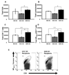
Figure 1. The frequency of cells, including antigen specific CD8+ T cells, was significantly increased in the lung of influenza infected mice that received SDR DCs prior to infection
Lungs were removed from influenza infected mice receiving no DCs, HCC DCs, or SDR DCs. The proportion of (A) total cells (*p < 0.01), (B) total lymphocytes (*p < 0.01), (C) total CD8+ T cells (p < 0.02), and (D–E) total NP366-74CD8+ T cells (*p < 0.001) detected at day 9 of influenza infection was affected by treatment with SDR DCs. Data represent three separate experiments, with n=4–7 per group, per experiment. (**p<0.01 in SDR vs. NO DC condition)
Figure 2
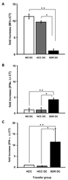
Figure 2. IFN-α and IFN-γ mRNA expression is increased, and M1 mRNA expression is decreased in the lung of influenza infected mice receiving SDR DCs prior to infection
The apical lobe of each lung was flash frozen and processed for PCR analysis of (A) M1 (*p < 0.01), (B) IFN-α (*p < 0.04), and (C) IFN-γ (*p < 0.05) mRNA expression. Data represent three separate experiments, with n=4–7 per group, per experiment. (**p < 0.5 in SDR vs. NO DC condition)
Figure 3
Figure 3. IFN-γ expression secretion by CD8+ Tcells is increased when antigen is presented by SDR DCs
CD11c+ DCs from SDR or HCC mice were purified and co-cultured with purified CD8+ T cells from influenza infected mice in a 1:5 ratio. Cultures were stimulated with 10µg/ml NP366-74 peptide and supernatants were harvested after 48 hours. Supernatants were analyzed by cytometric bead array, and only IFN-γ protein levels are shown, as differences in all other cytokines did not reach statistical significance. Data represent three separate experiments, with n=4–7 per group, per experiment. (*p < 0.05)
Figure 4
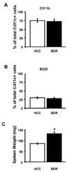
Figure 4. The majority of DCs in SDR and HCC mice are of a conventional phenotype
CD11c+ DCs were purified from SDR and HCC using Pan-DC microbeads (Miltenyi Biotec, Auburn, CA) and analyzed by flow cytometry for expression of CD11c, CD11b, and B220. Cells were gated on the monocyte population and analyzed for co-expression of (A) CD11b/CD11c or (B) B220/CD11c indicating a conventional or plasmacytoid phenotype, respectively. The size of the spleen of each animal in the SDR and HCC group was also assessed to confirm splenomegaly, indicative of that there was an SDR effect (C). Data represent three separate experiments, with n=4–7 per group, per experiment. (*p < 0.001)

Similar articles

Cited by

References

    1. Adler HS, Steinbrink K. Tolerogenic dendritic cells in health and disease: friend and foe! Eur J Dermatol. 2007;17:476–491. - PubMed
    1. Ali A, Avalos RT, Ponimaskin E, Nayak DP. Influenza virus assembly: effect of influenza virus glycoproteins on the membrane association of M1 protein. J Virol. 2000;74:8709–8719. - PMC - PubMed
    1. Angeli A, Masera RG, Sartori ML, Fortunati N, Racca S, Dovio A, Staurenghi A, Frairia R. Modulation by cytokines of glucocorticoid action. Ann N Y Acad Sci. 1999;876:210–220. - PubMed
    1. Avitsur R, Kavelaars A, Heijnen C, Sheridan JF. Social stress and the regulation of tumor necrosis factor-alpha secretion. Brain Behav Immun. 2005;19:311–317. - PubMed
    1. Bailey MT, Engler H, Powell ND, Padgett DA, Sheridan JF. Repeated social defeat increases the bactericidal activity of splenic macrophages through a Toll-like receptor-dependent pathway. Am J Physiol Regul Integr Comp Physiol. 2007;293:R1180–R1190. - PubMed

Publication types

MeSH terms