A cerebellar thalamic cortical circuit for error-related cognitive control
- PMID: 20656038
- PMCID: PMC2962720
- DOI: 10.1016/j.neuroimage.2010.07.042
A cerebellar thalamic cortical circuit for error-related cognitive control
Abstract
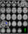
Error detection and behavioral adjustment are core components of cognitive control. Numerous studies have focused on the anterior cingulate cortex (ACC) as a critical locus of this executive function. Our previous work showed greater activation in the dorsal ACC and subcortical structures during error detection, and activation in the ventrolateral prefrontal cortex (VLPFC) during post-error slowing (PES) in a stop signal task (SST). However, the extent of error-related cortical or subcortical activation across subjects was not correlated with VLPFC activity during PES. So then, what causes VLPFC activation during PES? To address this question, we employed Granger causality mapping (GCM) and identified regions that Granger caused VLPFC activation in 54 adults performing the SST during fMRI. These brain regions, including the supplementary motor area (SMA), cerebellum, a pontine region, and medial thalamus, represent potential targets responding to errors in a way that could influence VLPFC activation. In confirmation of this hypothesis, the error-related activity of these regions correlated with VLPFC activation during PES, with the cerebellum showing the strongest association. The finding that cerebellar activation Granger causes prefrontal activity during behavioral adjustment supports a cerebellar function in cognitive control. Furthermore, multivariate GCA described the "flow of information" across these brain regions. Through connectivity with the thalamus and SMA, the cerebellum mediates error and post-error processing in accord with known anatomical projections. Taken together, these new findings highlight the role of the cerebello-thalamo-cortical pathway in an executive function that has heretofore largely been ascribed to the anterior cingulate-prefrontal cortical circuit.
Copyright © 2010 Elsevier Inc. All rights reserved.
Figures


References
-
- Abler B, Roebroeck A, Goebel R, Hose A, Schonfeldt-Lecuona C, Hole G, Walter H. Investigating directed influences between activated brain areas in a motor-response task using fMRI. Magn Reson Imaging. 2006;24:181–185. - PubMed
-
- Akaike H. A new look at the statistical model identification. Automatic Control, IEEE Transactions on. 1974;19:723.
-
- Allen G, Buxton RB, Wong EC, Courchesne E. Attentional activation of the cerebellum independent of motor involvement. Science. 1997;275:1940–1943. - PubMed
-
- Allen G, McColl R, Barnard H, Ringe WK, Fleckenstein J, Cullum CM. Magnetic resonance imaging of cerebellar-prefrontal and cerebellar-parietal functional connectivity. Neuroimage. 2005;28:39–48. - PubMed
-
- Allen GI, Tsukahara N. Cerebrocerebellar communication systems. Physiol Rev. 1974;54:957–1006. - PubMed
Publication types
MeSH terms
Grants and funding
LinkOut - more resources
Full Text Sources

