

The phosphatidylserine receptors, T cell immunoglobulin mucin proteins 3 and 4, are markers of histiocytic sarcoma and other histiocytic and dendritic cell neoplasms
- PMID: 20656318
- PMCID: PMC3115740
- DOI: 10.1016/j.humpath.2010.04.005
The phosphatidylserine receptors, T cell immunoglobulin mucin proteins 3 and 4, are markers of histiocytic sarcoma and other histiocytic and dendritic cell neoplasms
Abstract
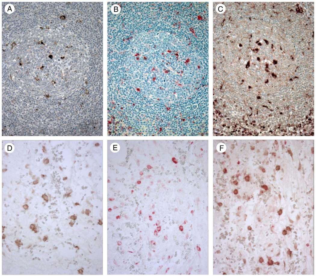
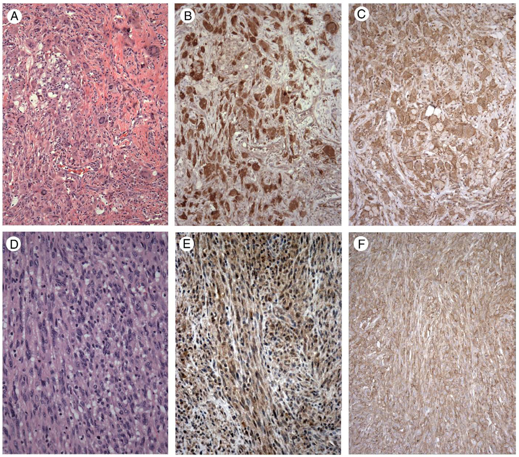
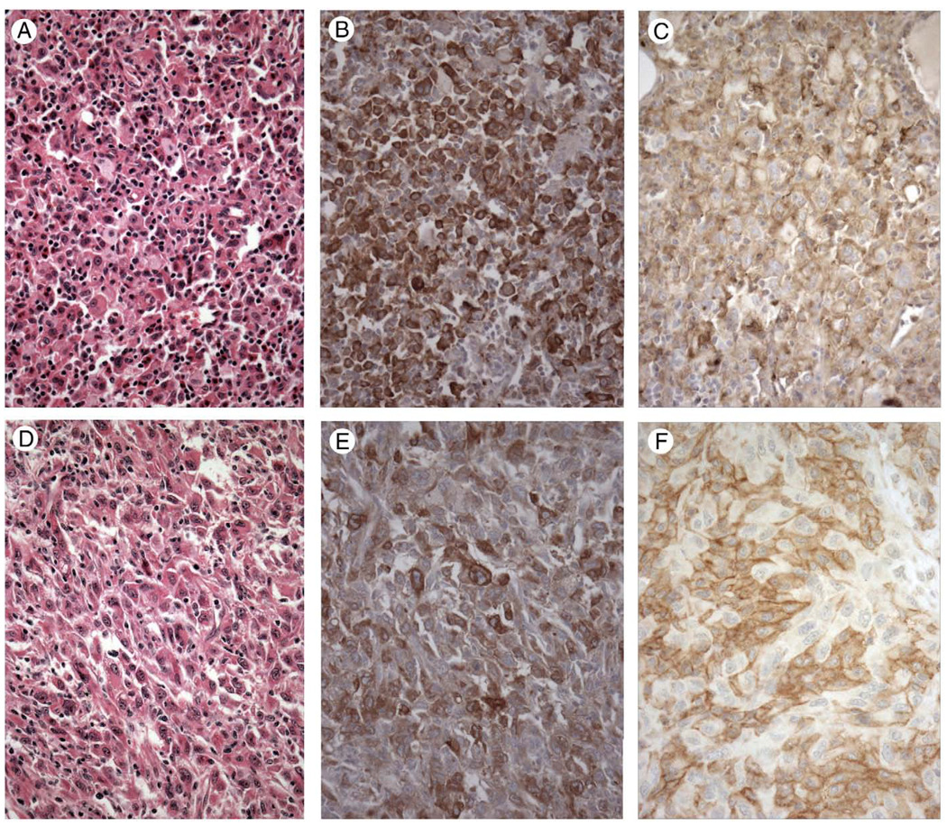
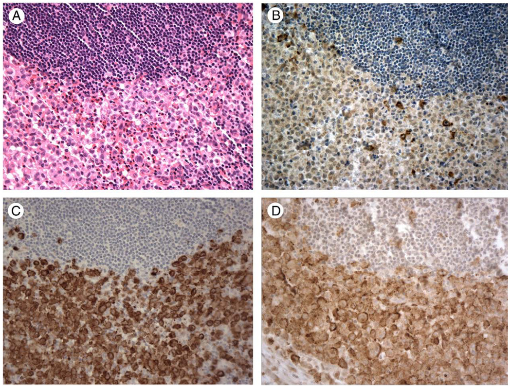
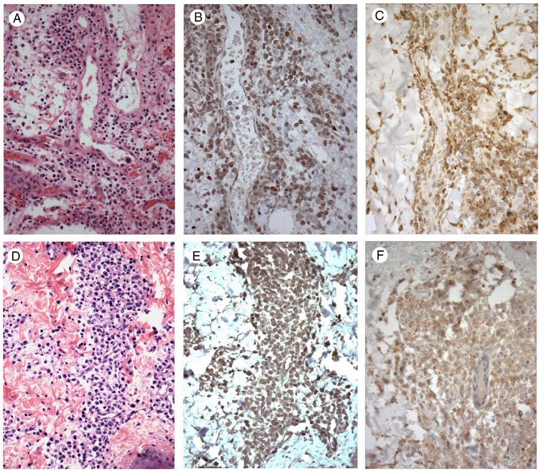
The T cell immunoglobulin mucin (TIM) proteins are a family of cell surface phosphatidyserine receptors that are important for the recognition and phagocytosis of apoptotic cells. Because TIM-4 is expressed by macrophages and dendritic cells in human tissue, we examined its expression in a range of histiocytic and dendritic cell neoplasms and found moderate to strong immunohistochemical staining in cases of juvenile xanthogranuloma and histiocytic sarcoma, and lower level staining in interdigitating dendritic cell sarcoma, Langerhans cell histiocytosis, acute monocytic leukemia (leukemia cutis), and blastic plasmacytoid dendritic cell neoplasm (hematodermic tumor). TIM-3 was first described on activated T(H)1 cells but was recently shown to also be a phosphatidylserine receptor and mediate phagocytosis. We found TIM-3 was expressed by peritoneal macrophages, monocytes and splenic dendritic cells. We found that it, like TIM-4, is expressed in a range of histiocytic and dendritic cell neoplasms, typically with strong immunohistochemical staining. Cases of diffuse large B cell lymphoma, anaplastic large cell lymphoma, metastatic malignant melanoma, and metastatic poorly differentiated carcinoma generally exhibited negative to minimal heterogenous staining for TIM-4 and TIM-3. We conclude that histiocytic and dendritic cell neoplasms consistently express TIM-3 and TIM-4 and that these molecules are new markers of neoplasms derived from histiocytic and dendritic cells.
Copyright © 2010 Elsevier Inc. All rights reserved.
Figures
Similar articles
-
Descriptive Analysis of Histiocytic and Dendritic Cell Neoplasms: A Single-Institution Experience.Yonsei Med J. 2020 Sep;61(9):774-779. doi: 10.3349/ymj.2020.61.9.774. Yonsei Med J. 2020. PMID: 32882761 Free PMC article.
-
High frequency of clonal IG and T-cell receptor gene rearrangements in histiocytic and dendritic cell neoplasms.Oncotarget. 2016 Nov 29;7(48):78355-78362. doi: 10.18632/oncotarget.13058. Oncotarget. 2016. PMID: 27823979 Free PMC article.
-
Histiocytic and dendritic cell neoplasms: Reappraisal of a Japanese series based on t(14;18) and neoplastic PD-L1 expression.Pathol Int. 2021 Jan;71(1):24-32. doi: 10.1111/pin.13044. Epub 2020 Nov 25. Pathol Int. 2021. PMID: 33238073
-
Histiocytic and Dendritic Cell Neoplasms.Surg Pathol Clin. 2019 Sep;12(3):805-829. doi: 10.1016/j.path.2019.03.013. Epub 2019 Jun 8. Surg Pathol Clin. 2019. PMID: 31352989 Review.
-
A review of histiocytic diseases of dogs and cats.Vet Pathol. 2014 Jan;51(1):167-84. doi: 10.1177/0300985813510413. Vet Pathol. 2014. PMID: 24395976 Review.
Cited by
-
TIM4+macrophages suppress the proinflammatory response to maintain the chronic alveolar echinococcosis infection.Front Cell Infect Microbiol. 2025 Jun 18;15:1600686. doi: 10.3389/fcimb.2025.1600686. eCollection 2025. Front Cell Infect Microbiol. 2025. PMID: 40606633 Free PMC article.
-
Glioma-derived T cell immunoglobulin- and mucin domain-containing molecule-4 (TIM4) contributes to tumor tolerance.J Biol Chem. 2011 Oct 21;286(42):36694-9. doi: 10.1074/jbc.M111.292540. Epub 2011 Sep 6. J Biol Chem. 2011. PMID: 21896488 Free PMC article.
-
T cell immunoglobulin- and mucin-domain-containing molecule 3 gene polymorphisms and susceptibility to pancreatic cancer.Mol Biol Rep. 2012 Nov;39(11):9941-6. doi: 10.1007/s11033-012-1862-y. Epub 2012 Jun 26. Mol Biol Rep. 2012. PMID: 22733499
-
Braf mutation in interdigitating dendritic cell sarcoma: a case report and review of the literature.Cancer Biol Ther. 2015;16(8):1128-35. doi: 10.1080/15384047.2015.1057359. Cancer Biol Ther. 2015. PMID: 26047060 Free PMC article. Review.
-
Immunological Targets for Immunotherapy: Inhibitory T Cell Receptors.Methods Mol Biol. 2020;2055:23-60. doi: 10.1007/978-1-4939-9773-2_2. Methods Mol Biol. 2020. PMID: 31502146 Free PMC article. Review.
References
-
- Nakayama M, AKiba H, Takeda K, et al. Tim-3 mediates phagocytosis of apoptotic cells and cross-presentation. Blood. 2009;113:3821–3830. - PubMed
Publication types
MeSH terms
Substances
Grants and funding
LinkOut - more resources
Full Text Sources
Other Literature Sources
Research Materials
