

Dynamic membrane remodeling at invadopodia differentiates invadopodia from podosomes
- PMID: 20656375
- PMCID: PMC3153956
- DOI: 10.1016/j.ejcb.2010.06.006
Dynamic membrane remodeling at invadopodia differentiates invadopodia from podosomes
Abstract
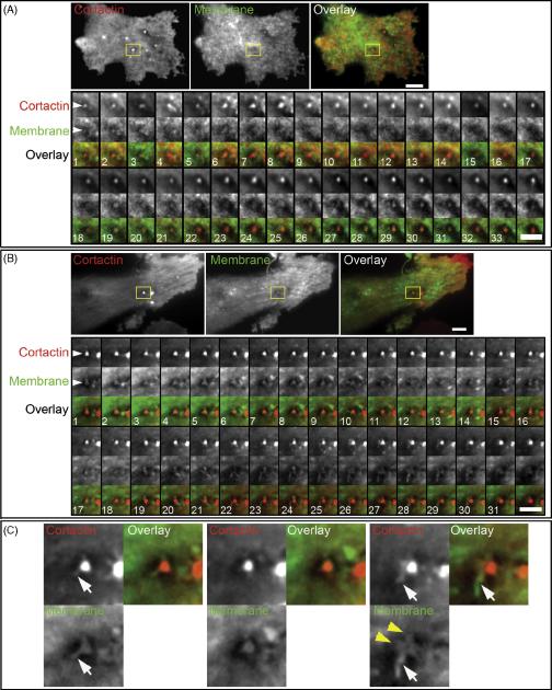
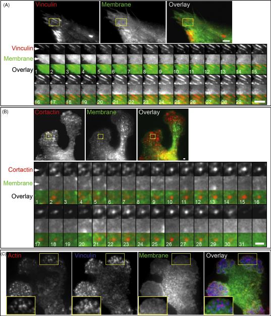
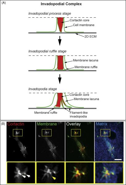
Invadopodia are specialized actin-rich protrusions of metastatic tumor and transformed cells with crucial functions in ECM degradation and invasion. Although early electron microscopy studies described invadopodia as long filament-like protrusions of the cell membrane adherent to the matrix, fluorescence microscopy studies have focused on invadopodia as actin-cortactin aggregates localized to areas of ECM degradation. The absence of a clear conceptual integration of these two descriptions of invadopodial structure has impeded understanding of the regulatory mechanisms that govern invadopodia. To determine the relationship between the membrane filaments identified by electron microscopy and the actin-cortactin aggregates of invadopodia, we applied rapid live-cell high-resolution TIRF microscopy to examine cell membrane dynamics at the cortactin core of the invadopodia of human carcinoma cells. We found that cortactin docking to the cell membrane adherent to 2D fibronectin matrix initiates invadopodium assembly associated with the formation of an invadopodial membrane process that extends from a ventral cell membrane lacuna toward the ECM. The tip of the invadopodial process flattens as it interacts with the 2D matrix, and it undergoes constant rapid ruffling and dynamic formation of filament-like protrusions as the invadopodium matures. To describe this newly discovered dynamic relationship between the actin-cortactin core and invadopodial membranes, we propose a model of the invadopodial complex. Using TIRF microscopy, we also established that - in striking contrast to the invadopodium - membrane at the podosome of a macrophage fails to form any process- or filament-like membrane protrusions. Thus, the undulation and ruffling of the invadopodial membrane together with the formation of dynamic filament-like extensions from the invadopodial cortactin core defines invadopodia as invasive superstructures that are distinct from the podosomes.
Copyright © 2010 Elsevier GmbH. All rights reserved.
Figures
References
-
- Abercrombie M, Heaysman JE, Pegrum SM. The locomotion of fibroblasts in culture. IV. Electron microscopy of the leading lamella. Exp Cell Res. 1971;67:359–367. - PubMed
-
- Akisaka T, Yoshida H, Suzuki R, Takama K. Adhesion structures and their cytoskeleton-membrane interactions at podosomes of osteoclasts in culture. Cell Tissue Res. 2008;331:625–641. - PubMed
-
- Akiyama SK. Curr Protoc Cell Biol. 2001. Purification of fibronectin; p. 15. Chapter 10, Unit 10. - PubMed
-
- Artym VV, Yamada KM, Mueller SC. ECM degradation assays for analyzing local cell invasion. Methods Mol Biol. 2009;522:211–219. - PubMed
-
- Artym VV, Zhang Y, Seillier-Moiseiwitsch F, Yamada KM, Mueller SC. Dynamic interactions of cortactin and membrane type 1 matrix metalloproteinase at invadopodia: defining the stages of invadopodia formation and function. Cancer Res. 2006;66:3034–3043. - PubMed
Publication types
MeSH terms
Substances
Grants and funding
LinkOut - more resources
Full Text Sources
Medical
Research Materials
Miscellaneous
