Comparative Study
. 2010 Sep;76(18):6171-9.
doi: 10.1128/AEM.01091-10. Epub 2010 Jul 23.

Quantitative PCR assay for Mycobacterium pseudoshottsii and Mycobacterium shottsii and application to environmental samples and fishes from the Chesapeake Bay

Affiliations
Comparative Study

Quantitative PCR assay for Mycobacterium pseudoshottsii and Mycobacterium shottsii and application to environmental samples and fishes from the Chesapeake Bay

D T Gauthier et al. Appl Environ Microbiol. 2010 Sep.

Abstract

Striped bass (Morone saxatilis) in the Chesapeake Bay are currently experiencing a very high prevalence of mycobacteriosis associated with newly described Mycobacterium species, Mycobacterium pseudoshottsii and M. shottsii. The ecology of these mycobacteria outside the striped bass host is currently unknown. In this work, we developed quantitative real-time PCR assays for M. pseudoshottsii and M. shottsii and applied these assays to DNA extracts from Chesapeake Bay water and sediment samples, as well as to tissues from two dominant prey of striped bass, Atlantic menhaden (Brevoortia tyrannus) and bay anchovy (Anchoa mitchilli). Mycobacterium pseudoshottsii was found to be ubiquitous in water samples from the main stem of the Chesapeake Bay and was also present in water and sediments from the Rappahannock River, Virginia. M. pseudoshottsii was also detected in menhaden and anchovy tissues. In contrast, M. shottsii was not detected in water, sediment, or prey fish tissues. In conjunction with its nonpigmented phenotype, which is frequently found in obligately pathogenic mycobacteria of humans, this pattern of occurrence suggests that M. shottsii may be an obligate pathogen of striped bass.


Figures

FIG. 1.
FIG. 1.
Dot blot hybridization of tester-specific fragment clones (spots) with AluI-digested, digoxigenin-labeled M. pseudoshottsii (Mp; left) or M. shottsii (Ms; right) genomic DNA. Blots are identical with respect to spotted DNA. Spots G4 and G5 (boxed) represent positive controls of M. pseudoshottsii and M. shottsii AluI-digested genomic DNA (unlabeled), respectively. Clone F5, the sequence of which was used to generate the M. shottsii primer/probe set used for assays, is circled.
FIG. 2.
FIG. 2.
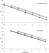
Standard curves generated from analysis of spiked nitrocellulose filters (solid lines and symbols) and sediment extractions (open symbols, dashed lines). Zero log dilution represents 1 × 10⁵ bacteria/reaction. Dilution series of tissue extracts are not shown for clarity, as CT values overlapped with those of water extractions. Duplicate CT values of results for triplicate tissue extractions at the zero dilution for M. pseudoshottsii were as follows: (i) 15.70, 15.63; (ii) 15.98, 16.13; (iii) 15.94, 16.16. Duplicate CT values of results for triplicate tissue extractions at the zero dilution for M. shottsii were as follows: (i) 21.29, 21.27; (ii) 21.55, 21.63; (iii) 21.35, 21.38. R2 values for all standard curves (including those using tissue) were >0.98.
FIG. 3.
FIG. 3.
Inhibition of qPCR assays for M. pseudoshottsii and M. shottsii by 250 ml water filtration residue or 200 mg sediment at various initial concentrations of target DNA. (A and C) Plots showing inhibition of qPCR detection of 10-fold dilutions of M. pseudoshottsii (A) and M. shottsii (C) in the presence of water residue (0.22 μm). (B and D) Plots showing inhibition due to sediment (200 mg) for M. pseudoshottsii (B) and M. shottsii (D). Data points for water are represented by solid symbols; data points for sediments are represented by open symbols. Standard curves (no residue/no sediment) are indicated by broken lines (−· ·−· ·−) and dotted lines (· · · · · · ·) for residue and sediment, respectively. Solid and dashed lines represent amplification in the presence of residue and sediment, respectively. Consistent with Fig. 2, log dilution of −1 represents 104 bacteria/reaction. Each reaction is in duplicate, and two dilution series are presented for each target-inhibitor combination. Sediment inhibition curves are each from a separate pool of three Rappahannock River sediment samples. These sediment pools were positive for M. pseudoshottsii; however, CT values exceeded 33 cycles in all cases.
FIG. 4.
FIG. 4.
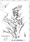
Densities of M. pseudoshottsii bacteria in the main stem of the Chesapeake Bay and the Rappahannock River (boxed) as measured by qPCR. Large circles represent sampling locations (surface water). Small circles (<0.4 M. pseudoshottsii/ml) represent positive samples with bacteria below the minimum quantitative threshold of the qPCR assay (see text).

Similar articles

Cited by

References

    1. Ablordey, A., M. Hilty, P. Stragier, J. Swings, and F. Portaels. 2005. Comparative nucleotide sequence analysis of polymorphic variable-number tandem-repeat loci in Mycobacterium ulcerans. J. Clin. Microbiol. 43:5281-5284. - PMC - PubMed
    1. Ablordey, A., J. Swings, C. Hubans, K. Chemlal, C. Locht, F. Portaels, and P. Supply. 2005. Multilocus variable-number tandem repeat typing of Mycobacterium ulcerans. J. Clin. Microbiol. 43:1546-1551. - PMC - PubMed
    1. Akopyants, N. S., A. Fradkov, L. Diatchenko, J. E. Hill, P. D. Siebert, S. A. Lukyanov, E. D. Sverdlov, and D. E. Berg. 1998. PCR-based subtractive hybridization and differences in gene content among strains of Helicobacter pylori. Proc. Natl. Acad. Sci. U. S. A. 95:13108-13113. - PMC - PubMed
    1. Blazer, V., W. K. Vogelbein, C. Densmore, D. Zwerner, and E. B. May. 1999. Ulcerative skin lesions in menhaden from Chesapeake Bay. J. Aquat. Anim. Health 11:340-349.
    1. Bruno, D. W., J. Griffiths, C. G. Mitchell, B. P. Wood, Z. J. Fletcher, F. A. Drobniewski, and T. S. Hastings. 1998. Pathology attributed to Mycobacterium chelonae infection among farmed and laboratory-infected Atlantic salmon Salmo salar. Dis. Aquat. Org. 33:101-109. - PubMed

Publication types

Associated data

LinkOut - more resources