. 2010 Oct;192(19):5071-80.
doi: 10.1128/JB.00575-10. Epub 2010 Jul 23.

Characterization of the PduS cobalamin reductase of Salmonella enterica and its role in the Pdu microcompartment

Affiliations

Characterization of the PduS cobalamin reductase of Salmonella enterica and its role in the Pdu microcompartment

Shouqiang Cheng et al. J Bacteriol. 2010 Oct.

Abstract

Salmonella enterica degrades 1,2-propanediol (1,2-PD) in a coenzyme B12 (adenosylcobalamin, AdoCbl)-dependent fashion. Salmonella obtains AdoCbl by assimilation of complex precursors, such as vitamin B12 and hydroxocobalamin. Assimilation of these compounds requires reduction of their central cobalt atom from Co3+ to Co2+ to Co+, followed by adenosylation to AdoCbl. In this work, the His6-tagged PduS cobalamin reductase from S. enterica was produced at high levels in Escherichia coli, purified, and characterized. The anaerobically purified enzyme reduced cob(III)alamin to cob(II)alamin at a rate of 42.3±3.2 μmol min(-1) mg(-1), and it reduced cob(II)alamin to cob(I)alamin at a rate of 54.5±4.2 nmol min(-1) mg(-1) protein. The apparent Km values of PduS-His6 were 10.1±0.7 μM for NADH and 67.5±8.2 μM for hydroxocobalamin in cob(III)alamin reduction. The apparent Km values for cob(II)alamin reduction were 27.5±2.4 μM with NADH as the substrate and 72.4±9.5 μM with cob(II)alamin as the substrate. High-performance liquid chromatography (HPLC) and mass spectrometry (MS) indicated that each monomer of PduS contained one molecule of noncovalently bound flavin mononucleotide (FMN). Genetic studies showed that a pduS deletion decreased the growth rate of Salmonella on 1,2-PD, supporting a role in cobalamin reduction in vivo. Further studies demonstrated that the PduS protein is a component of the Pdu microcompartments (MCPs) used for 1,2-PD degradation and that it interacts with the PduO adenosyltransferase, which catalyzes the terminal step of AdoCbl synthesis. These studies further characterize PduS, an unusual MCP-associated cobalamin reductase, and, in conjunction with prior results, indicate that the Pdu MCP encapsulates a complete cobalamin assimilation system.


Figures

FIG. 1.
FIG. 1.
Cobalamin assimilation and recycling pathway. Many organisms are able to take up CN-Cbl and OH-Cbl and convert them to the active coenzyme form, AdoCbl. This process involves reduction of the central cobalt atom of the corrin ring followed by addition of a 5′ deoxyadenosyl (Ado) group via a carbon-cobalt bond. The Ado group is unstable in vivo, and AdoCbl breaks down to form OH-Cbl. Consequently, cobalamin recycling is required for AdoCbl-dependent processes, and recycling uses the same pathway that functions in the assimilation of cobalamin from the environment. PPPi, triphosphate.
FIG. 2.
FIG. 2.
Sequence analyses of PduS. (A) Structure-based sequence alignment by Clustal X2 and PSIPRED. α-Helix and β-strand are represented by coil and arrow, respectively. CfPduS, PduS from Citrobacter freundii (GI 171854200); Ec, Escherichia coli (GI 157159374); Li, Listeria innocua (GI 16800175); Ri, Roseburia inulinivorans (GI 225376079); Se, Salmonella enterica (GI 16765383); Ye, Yersinia enterocolitica (GI 123442959); TaRnfC, RnfC from Thermanaerovibrio acidaminovorans (GI 269791702). (B) The domain architecture of the PduS protein (12). (C) Sequence logo of the amino acid residues (present at positions 25 to 39 in PduS of S. enterica) in the glycine-rich region used for nucleotide binding. Polar residues are in green, basic in blue, acidic in red, and hydrophobic in black.
FIG. 3.
FIG. 3.
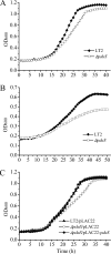
Phenotype and complementation of a pduS deletion mutation. Cells were grown in NCE minimal medium with 1,2-PD as the sole carbon and energy source. (A) The pduS deletion moderately impaired growth on 1,2-PD supplemented with 100 nM CN-Cbl. (B) The pduS deletion impaired growth and decreased cell density on 1,2-PD with limiting CN-Cbl (20 nM). (C) The pduS deletion was complemented by ectopic expression of pduS at 100 nM CN-Cbl. IPTG at 10 μM was used to induce production of PduS from pLAC22.
FIG. 4.
FIG. 4.
PduS is a component of the Pdu MCP. (A) Ten to 20% SDS-PAGE gel stained with Coomassie brilliant blue. Lane 1, molecular mass markers; lane 2, 10 μg Pdu MCPs purified from wild-type Salmonella. (B) Western blot for PduS. Lanes 1 and 2, 10 μg whole-cell extract or purified MCPs from the wild type; lanes 3 and 4, 10 μg whole-cell extract or purified MCPs from BE1352 (ΔpduS).
FIG. 5.
FIG. 5.
Proposed model for cobalamin recycling inside the Pdu MCPs of S. enterica. Prior studies showed that AdoCbl-dependent diol dehydratase (PduCDE) resides in the lumen of the Pdu MCP. PduCDE is subject to mechanism-based inactivation in which a damaged Cbl cofactor (OH-Cbl) becomes tightly enzyme bound. Diol dehydratase reactivase (PduGH) releases OH-Cbl from PduCDE. Then, OH-Cbl is reduced by PduS to cob(I)alamin and adenosylated to AdoCbl by the PduO adenosyltransferase. AdoCbl spontaneously associates with apo-PduCDE to form active holoenzyme. The recycling of OH-Cbl is required to maintain the activity of PduCDE and hence is essential for the degradation of 1,2-propanediol as a carbon and energy source. The finding that PduS is a component of the Pdu MCP suggests that cobalamin recycling can occur entirely within the MCP lumen.

Similar articles

Cited by

References

    1. Banerjee, R. 2006. B12 trafficking in mammals: a case for coenzyme escort service. ACS Chem. Biol. 1:149-159. - PubMed
    1. Banerjee, R. (ed.). 1999. Chemistry and Biochemistry of B12. John Wiley and Sons, New York, NY.
    1. Banerjee, R., C. Gherasim, and D. Padovani. 2009. The tinker, tailor, soldier in intracellular B12 trafficking. Curr. Opin. Chem. Biol. 13:484-491. - PMC - PubMed
    1. Berkowitz, D., J. M. Hushon, H. J. Whitfield, Jr., J. Roth, and B. N. Ames. 1968. Procedure for identifying nonsense mutations. J. Bacteriol. 96:215-220. - PMC - PubMed
    1. Blanche, F., M. Couder, L. Debussche, D. Thibaut, B. Cameron, and J. Crouzet. 1991. Biosynthesis of vitamin B12: stepwise amidation of carboxyl groups b, d, e, and g of cobyrinic acid a,c-diamide is catalyzed by one enzyme in Pseudomonas denitrificans. J. Bacteriol. 173:6046-6051. - PMC - PubMed

Publication types

MeSH terms

LinkOut - more resources