. 2010 Sep;9(9):1403-15.
doi: 10.1128/EC.00126-10. Epub 2010 Jul 23.

The Aspergillus fumigatus cspA gene encoding a repeat-rich cell wall protein is important for normal conidial cell wall architecture and interaction with host cells

Affiliations

The Aspergillus fumigatus cspA gene encoding a repeat-rich cell wall protein is important for normal conidial cell wall architecture and interaction with host cells

Emma Levdansky et al. Eukaryot Cell. 2010 Sep.

Abstract

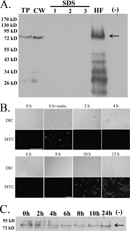
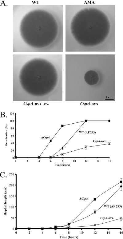
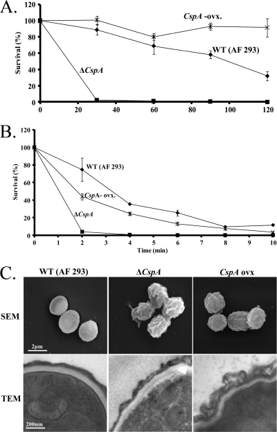
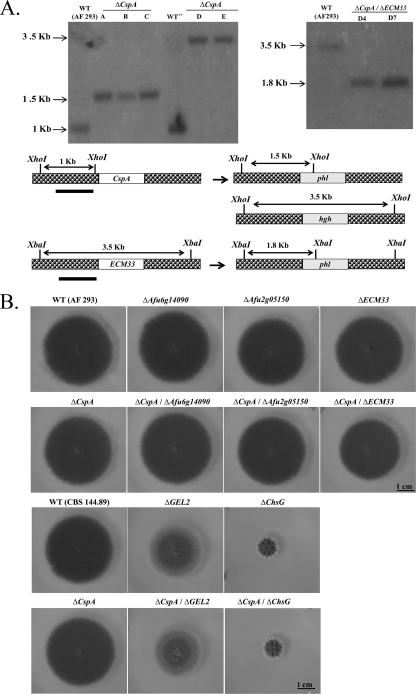
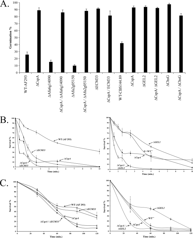
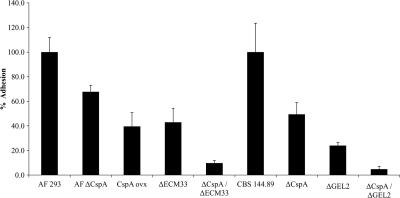
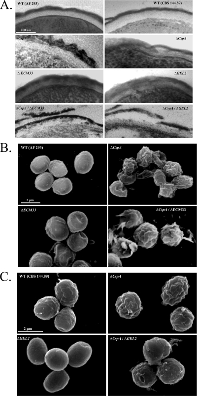
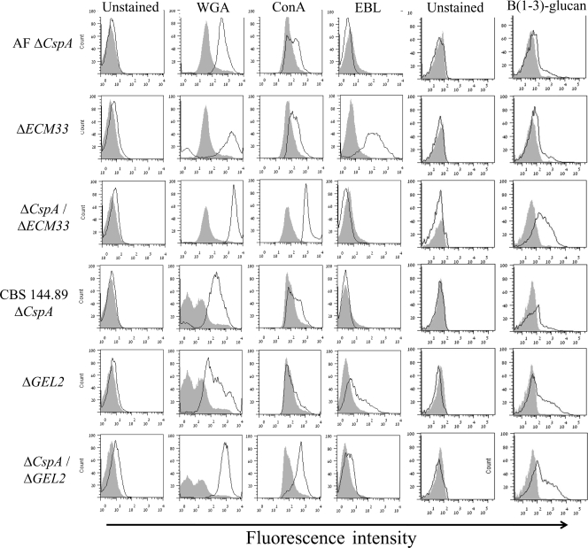
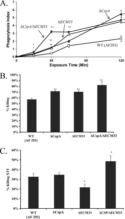
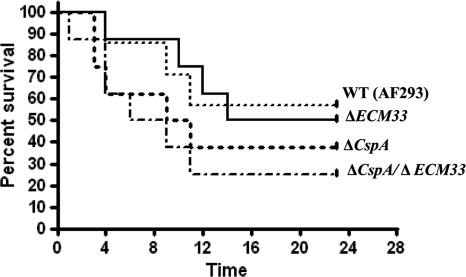
cspA (for cell surface protein A) encodes a repeat-rich glycophosphatidylinositol (GPI)-anchored cell wall protein (CWP) in the pathogenic fungus Aspergillus fumigatus. The number of repeats in cspA varies among isolates, and this trait is used for typing closely related strains of A. fumigatus. We have previously shown that deletion of cspA is associated with rapid conidial germination and reduced adhesion of dormant conidia. Here we show that cspA can be extracted with hydrofluoric acid (HF) from the cell wall, suggesting that it is a GPI-anchored CWP. The cspA-encoded CWP is unmasked during conidial germination and is surface expressed during hyphal growth. Deletion of cspA results in weakening of the conidial cell wall, whereas its overexpression increases conidial resistance to cell wall-degrading enzymes and inhibits conidial germination. Double mutant analysis indicates that cspA functionally interacts with the cell wall protein-encoding genes ECM33 and GEL2. Deletion of cspA together with ECM33 or GEL2 results in strongly reduced conidial adhesion, increased disorganization of the conidial cell wall, and exposure of the underlying layers of chitin and beta-glucan. This is correlated with increasing susceptibility of the DeltacspA, DeltaECM33, and DeltacspA DeltaECM33 mutants to conidial phagocytosis and killing by human macrophages and hyphal damage induced by neutrophils. However, these strains did not exhibit altered virulence in mice with infected lungs. Collectively, these results suggest a role for cspA in maintaining the strength and integrity of the cell wall.


Figures

Fig. 1.
Fig. 1.
Analysis of the protein encoded by cspA. (A) cspA-encoded protein is removed by HF treatment, suggesting that it is GPI anchored to the cell wall. Total protein (TP) and cell wall (CW) were extracted from the A. fumigatus myc-tagged cspA strain after 24 h of growth. CW was isolated and extracted three times with SDS/β-mercaptoethanol buffer (SDS lanes 1 to 3) and HF pyridine (lane HF). Total protein extracted from the untagged parental strain (AF 293) grown for 24 h was used as a negative control (−). myc-tagged cspA-encoded protein was detected with a specific mouse-derived anti-c-myc monoclonal antibody. Molecular mass markers (kDa) are indicated on the left side of the blot. (B) cspA-encoded protein is localized to the hyphal tip cell wall in germinating A. fumigatus conidia. Immunofluorescence analysis of cspA protein localization was performed with freshly harvested, nongerminated (0 h) conidia, sonicated conidia (0 h + sonic), and germlings (2 to 12 h) expressing cspA-myc. No immunofluorescence was detectable in nontransformed control WT cells (data not shown). Top panels (DIC), Cells viewed by differential interfering contrast microscopy; bottom panels (MYC), cspA-myc-tagged fluorescent images. Bars, 20 μm. (C) cspA protein is found in the cell wall of dormant and growing A. fumigatus conidia. Cell wall was extracted from nongerminated (0 h) conidia and germlings (2 to 12 h) expressing cspA-myc protein and analyzed by Western blotting.
Fig. 2.
Fig. 2.
cspA overexpression results in slow growth and reduced rates of germination and hyphal elongation. (A) WT (AF 293.1), AMA (AF 293.1 transformed with the AMA-1 control plasmid), cspA-ovx (overexpression strain), and cspA ovx-ev (the cspA-ovx strain following FOA-induced eviction of the AMA-1-CSP plasmid) strains were grown for 72 h on YAG agar. (B) Germination rates of the WT, cspA-deleted (ΔcspA), and cspA-ovx A. fumigatus strains. (C) Quantitative analysis of hyphal growth of WT, ΔcspA, and cspA-ovx A. fumigatus strains. The results for each time point are calculated as the mean ± standard error (error bar) for 50 hyphae (see Materials and Methods for details). The experiments shown in panels B and C were repeated three times with similar results.
Fig. 3.
Fig. 3.
cspA overexpression enhances conidial resistance to enzymatic cell wall removal and affects the morphology of the cell wall. Wild-type (WT), cspA-deleted (ΔcspA), and cspA-overexpressing (cspA-ovx) A. fumigatus strains were incubated in the presence of cell wall-degrading enzymes (A) or physically disrupted by agitation in the presence of glass beads (B) for various times. The ΔcspA strain exhibited enhanced sensitivity to cell wall-degrading enzymes and to physical disruption by glass beads, whereas the cspA-ovx strain was more resistant to the former. The experiments shown in panels A and B were repeated three times in triplicate with similar results. A representative experiment is shown. (C) Scanning electron microscopy (top) and transmission electron microscopy (bottom) of WT, ΔcspA, and cspA-ovx conidia show that cspA deletion and overexpression affect cell wall morphology and structure.
Fig. 4.
Fig. 4.
Generation of cspA double mutants in A. fumigatus. (A) Left, Southern blot analysis (top) and restriction map (bottom) of A. fumigatus strains after deletion of cspA by the hph (lanes A, B, and C) and phl (lanes D and E) selectable markers. Results are shown for A. fumigatus control AF 293 and CBS 144.89 wild-type strains and CBS 144.89 ΔcspA (lane A), ΔAfu6g14090 (lane B), ΔAfu2g05150 (lane C), ΔGEL2 (lane D), and ΔchsG (lane E) mutant strains. Genomic DNA was digested with XhoI and hybridized with 32P-labeled ΔcspA 5′ DNA probe (restriction map, filled black line), resulting in 1-kb fragments for the WT and 3.5-kb and 1.5-kb fragments for the ΔcspA hph-deleted and phl-deleted strains, respectively (see restriction map). Right, deletion of ECM33 by the phl cassette. Southern blot verification of the A. fumigatus control AF 293 WT strain (lane WT) and ECM33-deleted isolates (lanes D4 and D7) is shown. Genomic DNA was digested with XbaI and hybridized with 32P-labeled ECM33-5′ DNA probe, resulting in a 3.5-kb fragment for the WT and a 1.8-kb fragment for the ΔcspA phl-deleted strains, respectively (see restriction map). (B) WT and single and double mutant strains were spot inoculated on YAG agar and grown for 72 h. No differences in radial growth were detected between the WT (AF 293), the single-gene deletion mutants (ΔAfu6g14090, ΔAfu2g05150, ΔECM33, ΔcspA), and the double-gene deletion mutants (ΔcspA ΔAfu6g14090, ΔcspA ΔAfu2g05150, ΔcspA ΔECM33). Radial growth was reduced in the ΔGEL2 and ΔchsG mutant strains but was not further reduced by the additional deletion of cspAcspA ΔECM33 and ΔcspA ΔGEL2).
Fig. 5.
Fig. 5.
Germination and resistance of WT and cspA single- and double-deletion mutant strains to cell wall disruption. (A) Percentage of germinated cells of WT and cspA single- and double-deletion mutant strains after 8 h of growth in MM at 37°C. Shown are the means + standard deviations (error bars) for three independent experiments. (B) Resistance of the various strains to physical disruption by glass beads. ΔcspA ΔECM33 and ΔcspA ΔGEL2 strains are more susceptible to glass bead disruption than the respective single-gene deletion mutants and AF 293 (WT) and CBS 144.89 (WT**) strains. (C) Resistance of the various strains to cell wall-degrading enzymes. The ΔcspA ΔGEL2 strain is more sensitive to cell wall-degrading enzymes than the single-gene deletion mutant and WT strain (right), whereas the ΔcspA ΔECM33 strain is not (left). The experiments in panels B and C were repeated three times with similar results. A representative experiment is shown.
Fig. 6.
Fig. 6.
The cspA single- and double-deletion mutant strains show reduced conidial adhesion to extracellular matrix (ECM). The levels of adhesion of the ΔcspA, cspA-ovx, ΔECM33, ΔGEL2, ΔcspA ΔECM33, and ΔcspA ΔGEL2 strains to ECM were compared to that of the WT AF 293 and CBS 144.89 strains. Shown are the means + standard deviations (error bars) for three independent experiments. Each mutant strain exhibited significantly reduced adhesion relative to its parental WT strain (P < 0.005). The adhesion of each of the double mutant strains was significantly reduced in comparison to their parental single mutant strains (P < 0.0005).
Fig. 7.
Fig. 7.
cspA functionally interacts with ECM33 and GEL2. Results of transmission electron microscopy (A) and scanning electron microscopy of WT (AF 293 [B] and CBS 144.89 [C]), ΔcspA, ΔECM33, ΔGEL2, ΔcspA ΔECM33, and ΔcspA ΔGEL2 strains are shown. ΔcspA ΔECM33 and ΔcspA ΔGEL2 double mutants show an additive phenotype characterized by severe damage to the conidial cell wall morphology and structure.
Fig. 8.
Fig. 8.
The outer conidial cell walls of the ΔcspA ΔGEL2 and ΔcspA ΔECM33 mutant strains contain high levels of exposed chitin, mannose, and glucan. Conidia were stained with fluorescent lectins WGA, ConA, and EBL and anti-β-glucan-specific antibodies and analyzed by flow cytometry. The results are summarized in fluorescence frequency distribution histograms: the gray-filled histogram represents the relative signal of the corresponding WT conidia, whereas the unfilled black histogram represents the relative signal of mutant conidia (number of fungal cells versus relative fluorescence intensity expressed as arbitrary units on a logarithmic scale). A total of 10,000 cells were examined per sample in all experiments.
Fig. 9.
Fig. 9.
Increased uptake and killing of the ΔcspA, ΔECM33, and ΔcspA ΔECM33 mutant strains by human monocyte-derived macrophages (HuMoDMs) and enhanced hyphal damage by neutrophils (PMNLs). (A) Conidial endocytosis by HuMoDMs over time; (B) conidial killing by HuMoDMs after 2 h of coincubation; (C) hyphal damage by human neutrophils after 14 h of coincubation. Shown are the means + standard deviations (error bars) for three independent experiments. **, P value of <0.05 for each mutant strain relative to the WT (AF 293) strain.
Fig. 10.
Fig. 10.
Deletion of cspA, ECM33, or both cspA and ECM33 does not affect virulence in a neutropenic murine model of pulmonary aspergillosis. Cyclophosphamide/cortisone acetate-treated mice (8 per group) were infected intranasally with an inoculum of 2.5 × 10⁵ ΔcspA, ΔECM33, or ΔcspA ΔECM33 conidia/mouse. Percent survival was monitored throughout the 28-day study period. This experiment was repeated twice, with similar results.

Similar articles

Cited by

References

    1. Aimanianda V., Bayry J., Bozza S., Kniemeyer O., Perruccio K., Elluru S. R., Clavaud C., Paris S., Brakhage A. A., Kaveri S. V., Romani L., Latge J. P. 2009. Surface hydrophobin prevents immune recognition of airborne fungal spores. Nature 460:1117–1121 - PubMed
    1. Bainbridge B. W. 1971. Macromolecular composition and nuclear division during spore germination in Aspergillus nidulans. J. Gen. Microbiol. 66:319–325 - PubMed
    1. Balajee S. A., de Valk H. A., Lasker B. A., Meis J. F., Klaassen C. H. 2008. Utility of a microsatellite assay for identifying clonally related outbreak isolates of Aspergillus fumigatus. J. Microbiol. Methods 73:252–256 - PubMed
    1. Balajee S. A., Tay S. T., Lasker B. A., Hurst S. F., Rooney A. P. 2007. Characterization of a novel gene for strain typing reveals substructuring of Aspergillus fumigatus across North America. Eukaryot. Cell 6:1392–1399 - PMC - PubMed
    1. Chabane S., Sarfati J., Ibrahim-Granet O., Du C., Schmidt C., Mouyna I., Prevost M. C., Calderone R., Latge J. P. 2006. Glycosylphosphatidylinositol-anchored Ecm33p influences conidial cell wall biosynthesis in Aspergillus fumigatus. Appl. Environ. Microbiol. 72:3259–3267 - PMC - PubMed

Publication types

MeSH terms

Substances