Comparative Study
. 2010 Dec 15;182(12):1482-91.
doi: 10.1164/rccm.200912-1877OC. Epub 2010 Jul 23.

Chitin particles are multifaceted immune adjuvants

Affiliations
Comparative Study

Chitin particles are multifaceted immune adjuvants

Carla A Da Silva et al. Am J Respir Crit Care Med. .

Abstract

Rationale: Chitin is a ubiquitous polysaccharide in fungi, insects, allergens, and parasites that is released at sites of infection. Its role in the generation of tissue inflammation, however, is not fully understood.

Objectives: We hypothesized that chitin is an important adjuvant for adaptive immunity.

Methods: Mice were injected with a solution of ovalbumin and chitin.

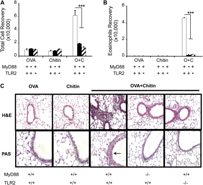
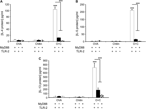
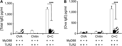
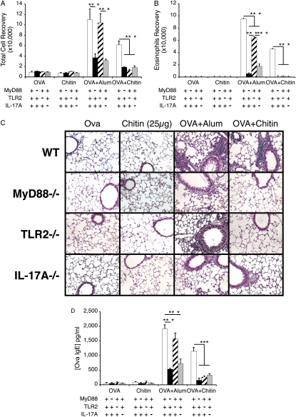
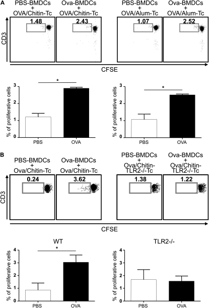
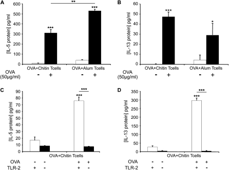
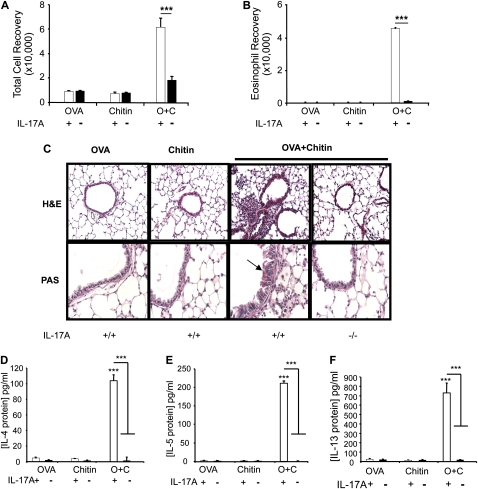
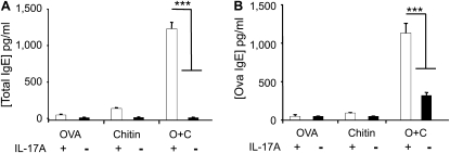
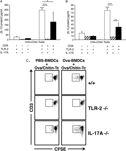
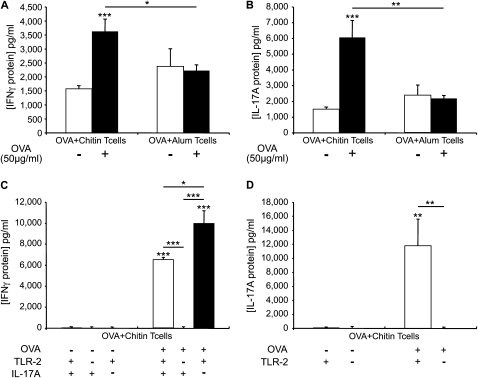
Measurements and main results: We used in vivo and ex vivo/in vitro approaches to characterize the ability of chitin fragments to foster adaptive immune responses against ovalbumin and compared these responses to those induced by aluminum hydroxide (alum). In vivo, ovalbumin challenge caused an eosinophil-rich pulmonary inflammatory response, Th2 cytokine elaboration, IgE induction, and mucus metaplasia in mice that had been sensitized with ovalbumin plus chitin or ovalbumin plus alum. Toll-like receptor-2, MyD88, and IL-17A played critical roles in the chitin-induced responses, and MyD88 and IL-17A played critical roles in the alum-induced responses. In vitro, CD4(+) T cells from mice sensitized with ovalbumin plus chitin were incubated with ovalbumin-stimulated bone marrow-derived dendritic cells. In these experiments, CD4(+) T-cell proliferation, IL-5, IL-13, IFN-γ, and IL-17A production were appreciated. Toll-like receptor-2, MyD88, and IL-17A played critical roles in these in vitro adjuvant properties of chitin. TLR-2 was required for cell proliferation, whereas IL-17 and TLR-2 were required for cytokine elaboration. IL-17A also inhibited the generation of adaptive Th1 responses.

Conclusions: These studies demonstrate that chitin is a potent multifaceted adjuvant that induces adaptive Th2, Th1, and Th17 immune responses. They also demonstrate that the adjuvant properties of chitin are mediated by a pathway(s) that involves and is regulated by TLR-2, MyD88, and IL-17A.


Figures

Figure 1.
Figure 1.
Adjuvant respiratory effects of chitin in vivo. MyD88 and Toll-like receptor 2 (TLR-2) sufficient (+/+; +) and deficient (−/−; −) mice received two intraperitoneal injections (Days 0 and 5) with a mixture containing ovalbumin (OVA) (20 μg), chitin (25 μg), OVA complexed with chitin, or its vehicle controls. One week later the mice received aerosolized OVA for 3 days, and 1 day after the last challenge the mice were killed, bronchoalveolar lavage (BAL) was undertaken, and lung sections were prepared. The effects of these treatments on (A) BAL total and (B) eosinophils recovery, and (C) tissue histology (hematoxylin and eosin and periodic acid-Schiff [PAS] stains, ×20 and ×40 of original magnification, respectively) were evaluated. Results are presented as the mean ± SEM of a minimum of six mice per group. ***P < 0.001. Arrow in C indicates PAS-positive airway mucus.
Figure 2.
Figure 2.
Adjuvant effects of chitin on bronchoalveolar lavage (BAL) Th2 cytokines in vivo. MyD88 and Toll-like receptor 2 (TLR-2) sufficient (+/+; +) and deficient (−/−; −) mice were sensitized at Day 0 and boosted at Day 5 with intraperitoneal injections containing ovalbumin (OVA) (20 μg), chitin (25 μg), OVA complexed with chitin, or their vehicle controls. One week later the mice received aerosolized OVA for 3 days, and 1 day after the last challenge the mice were killed and BAL was undertaken. Concentrations of (A) IL-4, (B) IL-5, and (C) IL-13 in BAL were measured by ELISA. Results are presented as the mean ± SEM of a minimum of six mice per group. ***P < 0.001.
Figure 3.
Figure 3.
Role of chitin in sensitization to ovalbumin (OVA) in vivo. MyD88 and Toll-like receptor 2 (TLR-2) sufficient (+/+; +) and deficient (−/−; −) mice were sensitized by two intraperitoneal injections (Days 0 and 5) of a mixture containing OVA (20 μg), chitin (25 μg), OVA complexed with chitin, or their vehicle controls. One week later the mice received aerosolized OVA for 3 days and 1 day after the last challenge were killed and serum was obtained. The levels of (A) total and (B) OVA-specific IgE were assessed by ELISA. Results are presented as the mean ± SEM of a minimum of six mice per group. ***P < 0.001.
Figure 4.
Figure 4.
Similarities and differences in chitin- and alum-induced in vivo responses. MyD88, Toll-like receptor 2 (TLR-2), and IL-17A sufficient (+/+; +) and deficient (−/−; −) mice received two intraperitoneal injections with a mixture containing ovalbumin (OVA) (20μg) plus chitin or alum or appropriate vehicle controls. One week later the mice received aerosolized OVA for 3 days, and 1 day after the last challenge the mice were killed. The effects of these treatments on (A) bronchoalveolar lavage (BAL) total and (B) eosinophils recovery, (C) tissue histology (hematoxylin and eosin stains, ×20 of original magnification), and (D) OVA-specific IgE were evaluated. Results in A, B, and D are presented as the mean ± SEM of a minimum of six mice per group. C is representative of three similar experiments. ***P < 0.001.
Figure 5.
Figure 5.
Effects of chitin in ovalbumin (OVA)-induced T-cell proliferation ex vivo. (A) Cocultures were prepared containing T cells (Tc) from wild-type (WT) mice immunized with OVA complexed to chitin (left pair) or alum (right pair) and OVA- or vehicle control–stimulated bone marrow–derived dendritic cells (BMDCs) from WT mice. (B) Identical cocultures were prepared using Tc from Toll-like receptor 2 (TLR-2)-sufficient and -deficient (TLR2−/−Tc) mice. Proliferation of carboxyfluorescein diacetate succinimidyl ester (CFSE)-labeled CD4+ T cells (vs. CD3 expression) was analyzed using flow cytometry at Day 5. Daughter cell generation is shown in the square box compared with undivided parental generation and is also represented as a percent of proliferative cells (histograms). One representative out of five independent experiments performed is shown. Results are presented as the mean ± SEM of a minimum of six mice per group. *P < 0.05.
Figure 6.
Figure 6.
Effects of chitin on ovalbumin (OVA)-induced cytokine production by T cells ex vivo. Cocultures were prepared that contained CD4+ T cells from wild-type (WT) mice (A and B) immunized with OVA complexed to chitin (OVA+Chitin T cells) or alum (OVA+ Alum T cells) and OVA (50 μg/ml; OVA+) or vehicle control–stimulated (OVA−) bone marrow–derived dendritic cells. Supernatants were collected after incubation for 3 days. CD4+ T cells from OVA/chitin-stimulated TLR-2 sufficient (+/+) and deficient (−/−) mice (C and D) were also used. The levels of IL-5 (A and C) and IL-13 (B and D) were measured by Bioplex ELISA. Results are presented as the mean ± SEM of a minimum of six mice per group. *P < 0.05, **P < 0.01, ***P < 0.001.
Figure 7.
Figure 7.
Roles of IL-17A in the adjuvant effects of chitin in vivo. Wild-type and IL-17A−/− mice were sensitized with OVA and chitin and then challenged with ovalbumin (OVA). (A) Total bronchoalveolar lavage (BAL) cell and (B) eosinophil recovery, (C) tissue inflammation (hematoxylin and eosin, ×20 final magnification), and mucus metaplasia (periodic acid-Schiff [PAS], ×40 final magnification), and the levels of (D) BAL IL-4, (E) IL-5, and (F) IL-13 were evaluated. Supernatant cytokines were measured using ELISA. The results are expressed as the mean ± SEM of a minimum of six mice per group. ***P < 0.001. The arrow in C indicates PAS-positive airway mucus.
Figure 8.
Figure 8.
Roles of IL-17A in the humoral adjuvant effects of chitin in vivo. Wild-type and IL-17A−/− mice were sensitized with ovalbumin (OVA) and chitin and then challenged with OVA. (A) Total and (B) OVA-specific IgE were evaluated. The results are expressed as the mean ± SEM of a minimum of six mice per group. ***P < 0.001.
Figure 9.
Figure 9.
Roles of IL-17A and Toll-like receptor 2 (TLR-2) in the in vitro adjuvant effects of chitin. Cocultures were prepared that contained CD4+ T cells from wild-type, IL-17A−/−, and TLR-2−/− mice immunized with ovalbumin (OVA) plus chitin (OVA+Chitin T cells) and OVA (50 μg/ml; OVA+) or vehicle control–stimulated (OVA−) bone marrow–derived dendritic cells (BMDCs). Supernatants were collected after incubation for 3 days and the levels of (A) IL-13 and (B) IL-5 were assessed by ELISA. (C) Proliferation of CFSE-labeled CD4+ T cells (vs. CD3 expression) was analyzed using flow cytometry at Day 5. Daughter cell generation is shown in the square box compared with undivided parental generation. Results in A and B are the mean ± SEM of a minimum of six mice per group. In C, one representative out of three independent experiments performed is shown. *P < 0.05, **P < 0.01, ***P < 0.001.
Figure 10.
Figure 10.
Roles of Toll-like receptor 2 (TLR-2) and IL-17A in ovalbumin (OVA) plus chitin-induced T cell IFN-γ and IL-17A production ex vivo. Cocultures were prepared that contained CD4+ T cells from wild-type mice (A and B) immunized with OVA complexed to chitin (OVA+Chitin T cells) or alum (OVA +Alum T cells) and OVA (50 μg/ml; OVA+) or vehicle control–stimulated (OVA−) bone marrow–derived dendritic cells. Supernatants were collected after incubation for 3 days. CD4+ T cells from OVA/chitin-stimulated TLR-2 sufficient (+/+) and deficient (−/−) and IL-17A sufficient (+/+) and deficient (−/−) mice (C and D) were also used. The levels of IFN-γ (A and C) and IL-17A (B and D) were assessed by ELISA. The results are expressed as the mean ± SEM of a minimum of six mice per group. *P < 0.05, **P < 0.01, ***P < 0.001.

References

    1. Neville AC, Parry DA, Woodhead-Galloway J. The chitin crystallite in arthropod cuticle. J Cell Sci 1976;21:73. - PubMed
    1. Fuhrman JA, Piessens WF. Chitin synthesis and sheath morphogenesis in Brugia malayi microfilariae. Mol Biochem Parasitol 1985;17:93. - PubMed
    1. Araujo AC, Souto-Padron T, de Souza W. Cytochemical localization of carbohydrate residues in microfilariae of Wuchereria bancrofti and Brugia malayi. J Histochem Cytochem 1993;41:571. - PubMed
    1. Debono M, Gordee RS. Antibiotics that inhibit fungal cell wall development. Annu Rev Microbiol 1994;48:471. - PubMed
    1. Zhu Z, Zheng T, Homer RJ, Kim YK, Chen NY, Cohn L, Hamid Q, Elias JA. Acidic mammalian chitinase in asthmatic Th2 inflammation and IL-13 pathway activation. Science 2004;304:1678. - PubMed

Publication types

MeSH terms