. 2010 Sep 24;285(39):30069-78.
doi: 10.1074/jbc.M110.148288. Epub 2010 Jul 23.

Metabolic regulator betaKlotho interacts with fibroblast growth factor receptor 4 (FGFR4) to induce apoptosis and inhibit tumor cell proliferation

Affiliations

Metabolic regulator betaKlotho interacts with fibroblast growth factor receptor 4 (FGFR4) to induce apoptosis and inhibit tumor cell proliferation

Yongde Luo et al. J Biol Chem. .

Abstract

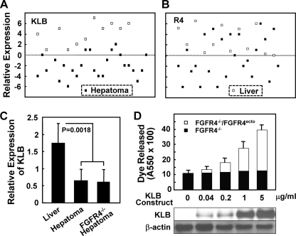
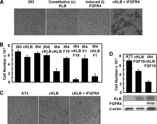
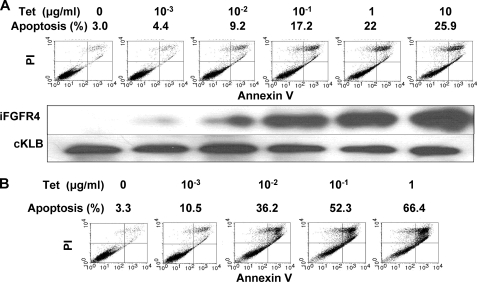
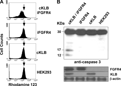
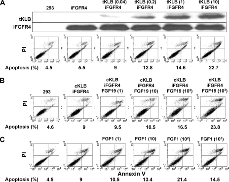
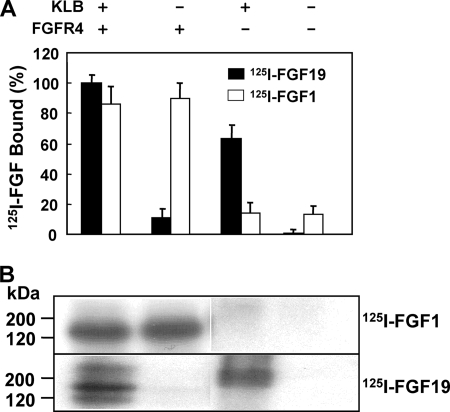
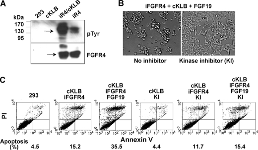
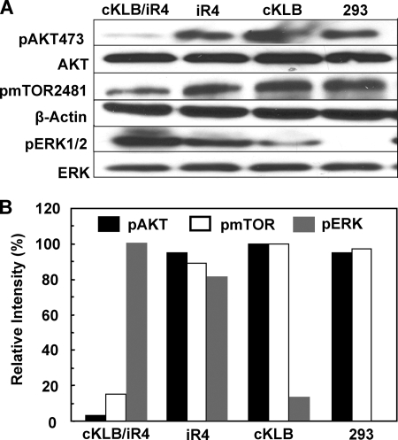
In organs involved in metabolic homeostasis, transmembrane α and βklothos direct FGFR signaling to control of metabolic pathways. Coordinate expression of βklotho and FGFR4 is a property of mature hepatocytes. Genetic deletion of FGFR4 or βklotho in mice disrupts hepatic cholesterol/bile acid and lipid metabolism. The deletion of FGFR4 has no effect on the proliferative response of hepatocytes after liver injury. However, its absence results in accelerated progression of dimethynitrosamine-initiated hepatocellular carcinomas, indicating that FGFR4 suppresses hepatoma proliferation. The mechanism underlying the FGFR4-mediated hepatoma suppression has not been addressed. Here we show that βklotho expression is more consistently down-regulated in human and mouse hepatomas than FGFR4. Co-expression and activation by either endocrine FGF19 or cellular FGF1 of the FGFR4 kinase in a complex with βklotho restricts cell population growth through induction of apoptotic cell death in both hepatic and nonhepatic cells. The βklotho-FGFR4 partnership caused a depression of activated AKT and mammalian target of rapamycin while activating ERK1/2 that may underlie the pro-apoptotic effect. Our results show that βklotho not only interacts with heparan sulfate-FGFR4 to form a complex with high affinity for endocrine FGF19 but also impacts the quality of downstream signaling and biological end points activated by either FGF19 or canonical FGF1. Thus the same βklotho-heparan sulfate-FGFR4 partnership that mediates endocrine control of hepatic metabolism plays a role in cellular homeostasis and hepatoma suppression through negative control of cell population growth mediated by pro-apoptotic signaling.


Figures

FIGURE 1.
FIGURE 1.
Reduced KLB expression in hepatomas and FGFR4-dependent effect of KLB expression in hepatoma cells. A and B, KLB and FGFR4 expression in human liver and hepatomas. Data from Affymetrix mRNA expression analyses of KLB and FGFR4 in human liver and clinically annotated hepatomas was extracted from the SIB-CleanEx Database and plotted numerically. Closed squares, human hepatoma samples; open squares, normal liver samples. C, reduced KLB expression in mouse hepatomas. The samples were taken from DEN-initiated hepatomas from normal and FGFR4-deficient (FGFR4−/−) mice as described (29). The relative expression of KLB mRNA was determined by quantitative PCR. The indicated data are the means ± S.D. from triplicate analyses of 10 normal liver and 12 hepatoma samples. D, FGFR4-dependent increase in cell death induced by expression of KLB in mouse hepatoma cells. KLB was transiently expressed by the addition of the indicated amounts of the KLB-pEF1a construct to hepatoma cells from FGFR4-deficient mice (FGFR4−/−) or cells (FGFR4−/−/FGFR4ecto) in which FGFR4 had been restored by stable transfection (29). Cell death was assessed by the uptake and release of APOPercentageTM dye. The indicated data are the means ± S.D. of three independent experiments with replicate analyses. FGFR4 expression levels (29) and the indicated KLB expression levels were assessed by immunoblotting of whole cell lysates using β-actin as the loading control.
FIGURE 2.
FIGURE 2.
Co-expression of KLB and FGFR4 also restricts nonhepatic cell population growth. A, cell morphology. Untransfected T-Rex-293 cells (293), cells stably transfected with KLB (cKLB), cells transfected with inducible FGFR4 cDNA (iFGFR4), and cKLB cells bearing inducible FGFR4 cDNA were examined by light microscopy after exposure overnight to 1 μg/ml Tet and 300 ng/ml of FGF19. B, population growth rates. The data are cell numbers after 5 days of culture. FGF19 or FGF1 was present at 300 ng/ml where indicated. The data are the means ± S.D. from three independent experiments. (R4), cells bearing inducible FGFR4 cDNA in Tet-free medium. C and D, co-expression of KLB and FGFR4 inhibits malignant prostate tumor cell growth. The induction of FGFR4 and effect on population dynamics of AT3 cells was determined as described for 293 cells. Expression of KLB and FGFR4 were measured by immunoblotting of whole cell lysates. β-Actin was used as the loading control.
FIGURE 3.
FIGURE 3.
KLB-dependent apoptosis is proportional to FGFR4 expression. A, apoptotic cell death after induction of FGFR4 for 24 h in 293 cells. FGFR4 was induced in KLB-expressing cells by the indicated concentrations of Tet. Apoptosis was monitored by flow cytometric analysis of membrane phospholipid exposure and propidium iodide uptake. Expression of FGFR4 and KLB were assessed by immunoblotting. B, rates of apoptosis in KLB-expressing 293 cells after induction of FGFR4 for 3 days.
FIGURE 4.
FIGURE 4.
Loss of mitochondrial transmembrane potential and induction of caspase cleavage by co-expression of KLB and FGFR4. A, mitochondrial membrane potential. Uptake and retention of the cationic voltage-sensitive lipophilic dye Rhodamine 123 in 293 cells expressing the indicated combinations of KLB and FGFR4 and treated with Tet and FGF19 were assessed by flow cytometry. B, cleavage of caspase 3. The lysates from 293 cells co-expressing KLB and FGFR4 after treatment with Tet and FGF19 were separated by SDS-PAGE, and the cleavage of caspase 3 into active fragments was then assessed by immunoblot analysis. The levels of FGFR4 and KLB were shown by immunoblotting of whole cell lysates as in Fig. 2.
FIGURE 5.
FIGURE 5.
FGFR4-dependent apoptotic cell death increases with increasing KLB expression and is stimulated by either FGF19 or FGF1. A, transient expression of KLB in FGFR4-expressing cells. 293 cells harboring inducible FGFR4 cDNA were transiently transfected (tKLB) overnight by the amount of KLB-pEF1a in μg/ml indicated in parentheses. FGFR4 was then induced by the addition of 10 ng/ml of Tet for 24 h followed by analysis of apoptosis and expression. B and C, stimulation of KLB-FGFR4-induced apoptosis by either FGF19 or FGF1. FGF19 (B) or FGF1 (C) was added at the concentrations in ng/ml indicated in parentheses to cells co-expressing constitutive KLB and FGFR4 induced by 10 ng/ml Tet.
FIGURE 6.
FIGURE 6.
Binding and complex formation among KLB, FGFR4, FGF19, and FGF1. A, differential binding of 125I-labeled FGF1 and FGF19. 293 cells expressing cKLB, iFGFR4, or their combination after Tet induction for 24 h were incubated with labeled FGF1 (white bar) or FGF19 (black bar). Cell surface bound radioactivity was determined by γ-counter. The data are the means ± S.D. of three independent experiments. B, covalent affinity cross-linked complexes. After binding with 10 ng/ml labeled FGF, the covalent affinity cross-linker disuccinimidyl suberate was used to cross-link the formed complex as described (37).
FIGURE 7.
FIGURE 7.
Activation and dependence of apoptotic cell death on activity of the FGFR4 tyrosine kinase. A, activation of the FGFR4 kinase by KLB. 293 cells co-expressing constitutive KLB and FGFR4 induced by 300 ng/ml Tet overnight were maintained in serum-free medium for 6 h (33) and then exposed to 300 ng/ml of FGF19 for 10 min followed by immunoblot analysis of lysates with anti-phosphotyrosine (pTyr) and anti-FGFR4 antibodies. B and C, rescue of KLB-FGFR4-induced apoptosis and cell death by inhibition of tyrosine kinase activity. The FGFR kinase inhibitor 1-(2-amino-6-(3,5-dimethoxyphenyl) pyrido [2,3-d] pyrimidin-7-yl)-3-tert-butyl urea (341608; Calbiochem, San Diego, CA) at 1 μm was added during the 24-h induction of FGFR4 by 300 ng/ml Tet in cells expressing KLB followed by analysis of cell morphology (B) and apoptosis and cell death (C). FGF19 was present at 1 μg/ml.
FIGURE 8.
FIGURE 8.
Selective depression of AKT and mTOR activity by the KLB-FGFR4 partnership. A, 293 cells were treated with 300 ng/ml Tet overnight followed by addition of 1 μg/ml FGF19 for 6 h and analysis of lysates by immunoblot with the indicated antibodies. B, quantification of pAKT, pmTOR, and pERK. The relative levels of pAKT to total AKT, pmTOR to β-actin, and pERK to total ERK were calculated from a densitometric scan of band intensities in A. 100% was the most intense band from each protein among samples.

References

    1. McKeehan W. L., Wang F., Kan M. (1998) Prog. Nucleic Acid Res. Mol. Biol. 59, 135–176 - PubMed
    1. Eswarakumar V. P., Lax I., Schlessinger J. (2005) Cytokine Growth Factor Rev. 16, 139–149 - PubMed
    1. Luo Y., Ye S., Kan M., McKeehan W. L. (2006) J. Biol. Chem. 281, 21052–21061 - PubMed
    1. Turner N., Grose R. (2010) Nat. Rev. Cancer. 10, 116–129 - PubMed
    1. Shimada T., Kakitani M., Yamazaki Y., Hasegawa H., Takeuchi Y., Fujita T., Fukumoto S., Tomizuka K., Yamashita T. (2004) J. Clin. Invest. 113, 561–568 - PMC - PubMed

Publication types

MeSH terms

Substances