Noninterleaved velocity encodings for improved temporal and spatial resolution in phase-contrast magnetic resonance imaging
- PMID: 20657227
- PMCID: PMC3071154
- DOI: 10.1097/RCT.0b013e3181d564e8
Noninterleaved velocity encodings for improved temporal and spatial resolution in phase-contrast magnetic resonance imaging
Abstract
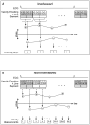
A segmented k-space acquisition technique using noninterleaved velocity encodings is presented to reduce spatial and temporal blur in phase-contrast cardiovascular magnetic resonance imaging. A translating phantom with pulsatile flow was used to simulate imaging of coronary arteries on a 1.5-T GE Echospeed scanner, using both interleaved and noninterleaved velocity encodings. The results demonstrate that the use of noninterleaved velocity encodings reduces spatial and temporal blur by improving the temporal resolution.
Figures


Similar articles
-
A Bayesian model for highly accelerated phase-contrast MRI.Magn Reson Med. 2016 Aug;76(2):689-701. doi: 10.1002/mrm.25904. Epub 2015 Oct 7. Magn Reson Med. 2016. PMID: 26444911 Free PMC article.
-
The application of breath hold phase velocity mapping techniques to the measurement of coronary artery blood flow velocity: phantom data and initial in vivo results.Magn Reson Med. 1994 May;31(5):526-36. doi: 10.1002/mrm.1910310509. Magn Reson Med. 1994. PMID: 8015406
-
Combined analysis of spatial and velocity displacement artifacts in phase contrast measurements of complex flows.J Magn Reson Imaging. 1997 Mar-Apr;7(2):339-46. doi: 10.1002/jmri.1880070214. J Magn Reson Imaging. 1997. PMID: 9090588
-
High efficiency multishot interleaved spiral-in/out: acquisition for high-resolution BOLD fMRI.Magn Reson Med. 2013 Aug;70(2):420-8. doi: 10.1002/mrm.24476. Epub 2012 Sep 28. Magn Reson Med. 2013. PMID: 23023395 Free PMC article.
-
Cardiovascular magnetic resonance phase contrast imaging.J Cardiovasc Magn Reson. 2015 Aug 9;17(1):71. doi: 10.1186/s12968-015-0172-7. J Cardiovasc Magn Reson. 2015. PMID: 26254979 Free PMC article. Review.
Cited by
-
Novel insight into the detailed myocardial motion and deformation of the rodent heart using high-resolution phase contrast cardiovascular magnetic resonance.J Cardiovasc Magn Reson. 2013 Sep 14;15(1):82. doi: 10.1186/1532-429X-15-82. J Cardiovasc Magn Reson. 2013. PMID: 24034168 Free PMC article.
References
-
- Hundley WG, Lange RA, Clarke GD, et al. Assessment of coronary arterial flow and flow reserve in humans with magnetic resonance imaging. Circulation. 1996 April 15;93(8):1502–8. - PubMed
-
- Davis CP, Liu PF, Hauser M, Gohde SC, von Schulthess GK, Debatin JF. Coronary flow and coronary flow reserve measurements in humans with breath-held magnetic resonance phase contrast velocity mapping. Magn Reson Med. 1997 April;37(4):537–44. - PubMed
-
- Hofman MB, Wickline SA, Lorenz CH. Quantification of in-plane motion of the coronary arteries during the cardiac cycle: implications for acquisition window duration for MR flow quantification. J Magn Reson Imaging. 1998 May;8(3):568–76. - PubMed
-
- Moran PR. A flow velocity zeugmatographic interlace for NMR imaging in humans. Magn Reson Imaging. 1982;1(4):197–203. - PubMed
-
- Haacke EM, Bearden FH, Clayton JR, Linga NR. Reduction of MR imaging time by the hybrid fast-scan technique. Radiology. 1986 February;158(2):521–9. - PubMed
Publication types
MeSH terms
Grants and funding
LinkOut - more resources
Full Text Sources
Medical

