Review
. 2010 Apr 30;15(5):3228-59.
doi: 10.3390/molecules15053228.

Vitamin B12: unique metalorganic compounds and the most complex vitamins

Affiliations
Review

Vitamin B12: unique metalorganic compounds and the most complex vitamins

Lucio Randaccio et al. Molecules. .

Abstract

The chemistry and biochemistry of the vitamin B(12) compounds (cobalamins, XCbl) are described, with particular emphasis on their structural aspects and their relationships with properties and function. A brief history of B(12), reveals how much the effort of chemists, biochemists and crystallographers have contributed in the past to understand the basic properties of this very complex vitamin. The properties of the two cobalamins, the two important B(12) cofactors Ado- and MeCbl are described, with particular emphasis on how the Co-C bond cleavage is involved in the enzymatic mechanisms. The main structural features of cobalamins are described, with particular reference to the axial fragment. The structure/property relationships in cobalamins are summarized. The recent studies on base-off/base-on equilibrium are emphasized for their relevance to the mode of binding of the cofactor to the protein scaffold. The absorption, transport and cellular uptake of cobalamins and the structure of the B(12) transport proteins, IF and TC, in mammals are reviewed. The B(12) transport in bacteria and the structure of the so far determined proteins are briefly described. The currently accepted mechanisms for the catalytic cycles of the AdoCbl and MeCbl enzymes are reported. The structure and function of B(12) enzymes, particularly the important mammalian enzymes methyltransferase (MetH) and methyl-malonyl-coenzyme A mutase (MMCM), are described and briefly discussed. Since fast proliferating cells require higher amount of vitamin B(12) than that required by normal cells, the study of B(12 )conjugates as targeting agents has recently gained importance. Bioconjugates have been studied as potential agents for delivering radioisotopes and NMR probes or as various cytotoxic agents towards cancer cells in humans and the most recent studies are described. Specifically, functionalized bioconjugates are used as "Trojan horses" to carry into the cell the appropriate antitumour or diagnostic label. Possible future developments of B(12) work are summarized.


Figures

Figure 1
Figure 1
(a) Structural formulas of vitamin B12 (X=CN) and of the biologically active cobalamins (X=Ado, X=CH3); (b) conventional atoms nomenclature for cobalamins; (c) base-on form (left side) and base-off form (right side); only the number or the symbol are indicated for the carbon atoms.
Figure 2
Figure 2
a) The main four resonance structures for the delocalized corrin system; b) mean bond lengths within the delocalised moiety of the corrin nucleus [25]; c) the folding angle ϕ. The double blue line represents the trace of the 5,6-benzimidazole group.
Scheme 1
Scheme 1
Dissociation equilibrium of the benzimidazole ligand to give the unprotonated (1) and protonate (2) base-off form of cobalamin.
Figure 3
Figure 3
Absorption, transport and cellular uptake of cobalamins in mammals. The schematic formation and function of the two cofactors, AdoCbl and MeCbl within the cell. The homolytic and heterolytic cleavage of the Co-C bond in AdoCbl and MeCbl enzymes, respectively are evidenced. The corresponding arrow indicates where the cleavage occurs, homolysis into the mitochondrion and heterolysis into the cytoplasm.
Figure 4
Figure 4
The α-β domain structure of transcobalamin (TC) and human intrinsic factor (IF). a) The two-domain structure of TC; the internal six α helices are in red and the six external ones in violet. The β domain is in blue. The loop connecting the two domains is in green. b) The two-domain structure of IF; the internal six α helices are in red and the six external ones in violet. The β domain is in blue. The figures are generated from PDB files 2BB5 (a) and 2PMV (b).
Figure 5
Figure 5
Hypothetical scheme of cobalamin uptake in E. coli. The proteins involved in the transport and B12 cellular uptake, whose X-ray structures are available, are shown by the ribbon representation for the protein moiety and by stick and ball for cobalamin. The X-ray structures of OmpF, ExbB and ExbD have not been so far determined and are indicated by a coloured form positioned into the membranes. The figures of proteins are generated from PDB files 2GUF (BtuB), 2GSK (BtuB-TonB), 1N2Z (BtuF) and 2QI9 (BtuC-BtuD-BtuF).
Figure 6
Figure 6
The three module representation of cobalamin methyltransferases.
Figure 7
Figure 7
The catalytic cycle and the coenzyme reactivation in methionine synthase. Reactions 1 and 2 are involved in the catalytic cycle and reaction 3 in the cobalamin reactivation. In the reactivation reaction, it has been suggested that histidine is displaced from cobalt in cob(II)alamin as shown on the top of the drawing.
Figure 8
Figure 8
Proposed four conformational states of MetH in solution: (a) unknown conformation in the rest state; (b) conformation for reaction 1 in Figure 7; (c) conformation for reaction 2 in Figure 7; (d) conformation for reaction 3 in Figure 7. The four modules are shown in green (Hcy), in blue (Fol), in gray (Cap-Cob) and in magenta (Adomet) respectively. The red rectangle represents the cobalamin and the vertical line in a), (b) and (c) represents its binding to the histidine of the Cob domain. In d) the position of the vertical line indicates that the histidine is displaced from Co.
Figure 9
Figure 9
The Rossmann fold found in: (a) Fol domain (PDB 1Q8J); (b) Cob domain of (PDB 3BUL) MetH from Escherichia coli. The His domain is similar to that of Fol.
Figure 10
Figure 10
The large movement of the Cap sub-domain of methionine synthase from the inactive Cap-Cob domain (in silver cartoon, PDB 1K7Y) to the active form (gold cartoon, PDB 1BMT). The Cob sub-domain with bonded cobalamin (in stick) is shown by red cartoons.
Figure 11
Figure 11
Ribbon representation of CoFeSP from C. hydrogenoformans (PDB 2H9A). The CfsB domain is shown in yellow cartoon and CfsA in silver cartoon. Cobalamin and iron-sulphur cluster are shown as sticks.
Figure 12
Figure 12
Ribbon representation of Methylmalonyl-CoA Mutase, MMCA (PDB 7REQ). The monomers α and β are shown in yellow and silver cartoon, respectively. Cobalamin is shown by orange sticks, while 2-carboxypropyl-CoA is represented with green sticks.
Figure 13
Figure 13
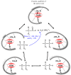
The catalytic cycle in AdoCbl enzymes (CH2R = Ado). The blue arrow indicates the final product in eliminases. The broken lines into the active site represent a sketch of the H-bonds between the cofactor and the protein aminoacid residues.
Figure 14
Figure 14
Sketch of a B12 bioconjugate and its functioning for imaging (step 1 and 2) and for antitumour activity (step 2). In step 1 the inactive labelled complex enters the tumour cell. In step 2 the drug is released by cleavage of its binding to the linker, by different way (pH, enzymatic process, etc.).
Figure 15
Figure 15
Some more recent B12 bioconjugates.

References

    1. Folkers K. History of Vitamin B12: Pernicious Anemia to Crystalline Cyanocobalamin. In: Dolphin D., editor. Vitamin B12. Volume I. John Wiley & Sons; New York, NY, USA: 1982. pp. 1–15.
    1. Hodgkin D.C. New and Old Problem in the Structure Analysis of Vitamin B12. In: Zagalak B., Friedrich W., editors. Vitamin B12, Proceedings of the Third European Symposium on Vitamin B12 and Intrinsic Factors, Zurich, Switzerland, March 1999. Walter de Gruyter; Berlin, Germany: 1979. pp. 19–36.
    1. Woodward R.B. Synthetic vitamin B12. In: Zagalak B., Friedrich W., editors. Vitamin B12, Proceedings of the Third European Symposium on Vitamin B12 and Intrinsic Factors, Zurich, Switzerland, March 1999. Walter de Gruyter; Berlin: Germany; 1979. pp. 37–88.
    1. Kräutler B. In: Vitamin B12 and B12 Proteins. Kräutler B., Arigoni D., Golding B.T., editors. Wiley-VCH; Weinheim, Germany: 1998. pp. 3–43.
    1. Hogenkamp H.P.C. In: Chemistry and Biochemistry of B12. Banerjee R., editor. John Wiley & Sons; New York, NY, USA: 1999. pp. 3–8.

Publication types

LinkOut - more resources