. 2010 Aug;42(8):676-83.
doi: 10.1038/ng.629. Epub 2010 Jul 25.

Matriptase initiates activation of epidermal pro-kallikrein and disease onset in a mouse model of Netherton syndrome

Affiliations

Matriptase initiates activation of epidermal pro-kallikrein and disease onset in a mouse model of Netherton syndrome

Katiuchia Uzzun Sales et al. Nat Genet. 2010 Aug.

Abstract

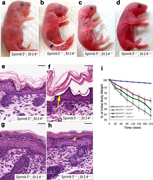
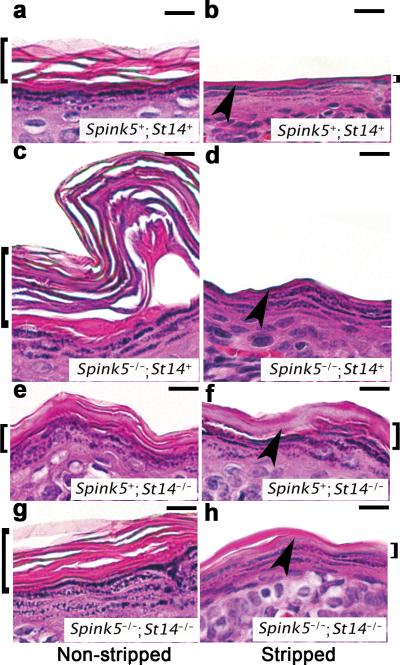
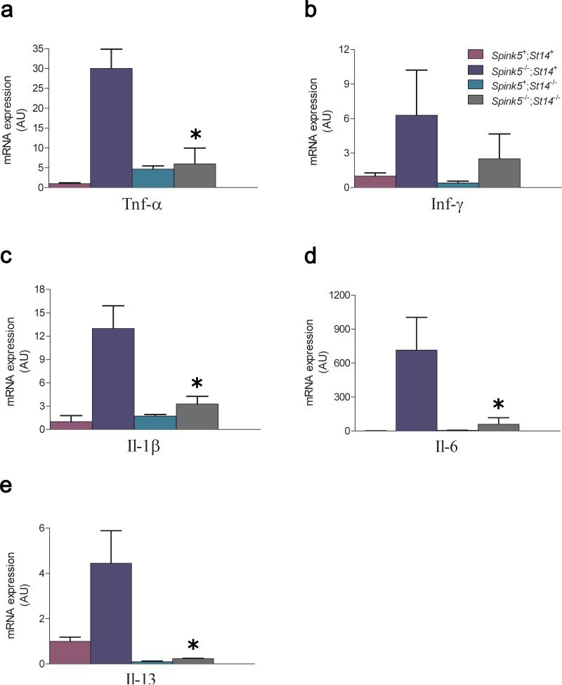
Deficiency in the serine protease inhibitor LEKTI is the etiological origin of Netherton syndrome, which causes detachment of the stratum corneum and chronic inflammation. Here we show that the membrane protease matriptase initiates Netherton syndrome in a LEKTI-deficient mouse model by premature activation of a pro-kallikrein cascade. Auto-activation of pro-inflammatory pro-kallikrein-related peptidases that are associated with stratum corneum detachment was either low or undetectable, but they were efficiently activated by matriptase. Ablation of matriptase from LEKTI-deficient mice dampened inflammation, eliminated aberrant protease activity, prevented detachment of the stratum corneum, and improved the barrier function of the epidermis. These results uncover a pathogenic matriptase-pro-kallikrein pathway that could operate in several human skin and inflammatory diseases.


Figures

Figure 1
Figure 1. Matriptase and LEKTI co-localize at the site of epidermal separation in Netherton syndrome
X-gal staining (cyan) (a), LEKTI immunostaining (brown) (b), and combined X-gal staining and immunohistochemistry (c) of epidermal sections from mice carrying a β-galactosidase-tagged St14 allele. Matriptase and LEKTI demonstrate co-localization at the granular-transitional layer (arrowheads in a and c) where premature corneodesmosome degradation causes epidermal separation in Netherton syndrome. Staining specificity is shown by the absence of X-gal staining in an epidermal section from a wildtype mouse (d) and lack of LEKTI immunoreactivity in a control section from a Spink5-/- mouse (e). Size bars all frames, 50 μm.
Figure 2
Figure 2. Activation of epidermal pro-kallikreins by matriptase
(a) Recombinant pro-kallikrein-related peptidase 5 (ProKLK5) (lanes 1 and 2) and pro-kallikrein-related peptidase 7 (proKLK7) (lanes 3 and 4) with C-terminal His tags were analyzed by Western blot using poly-histidine antibodies. Pro-kallikrein-related peptidase 5 appears as a single band with the predicted molecular weight of 40 kDa prior to deglycosylation (lane 1) and 32 kDa after deglycosylation (lane 2). Pro-kallikrein-related peptidase 7 appears as two bands of 28 and 34 kDa prior to deglycosylation (lane 3), and as a single band with the predicted 28 kDa molecular weight after deglycosylation (lane 4). Molecular weight standards (kDa) at left. b and c. Pro-kallikrein-related peptidase 5 and pro-kallikrein-related peptidase 7 (50 nM) were incubated for 15 min (b) or 30 min (c) with and without 2.5 nM matriptase. Fluorescence development was recorded after addition of a fluorogenic kallikrein-selective peptide. Reactions containing pro-kallikreins alone displayed minimal hydrolytic activity (brown and blue lines), compared to reactions containing pro-kallikreins and matriptase (red and black lines). d and e. Identical activation assays were performed substituting 2.5 nM matriptase with 2.5 nM kallkrein-related peptidase 5, demonstrating that pro-kallikrein-related peptidase 5, once converted to kallikrein-related peptidase 5 can activate pro-kallikrein-related peptidase 5 and pro-kallikrein-related peptidase 7. The contribution of matriptase in b and c and kallikrein-related peptidase 5 in d and e to fluorescence emission was subtracted for clarity. Experiments were performed in triplicate. Data are shown as mean fluorescence ± standard error of the mean.
Figure 3
Figure 3. Matriptase and prostasin are not inhibitory targets for LEKTI
(a) Recombinant GST (lanes 1 and 8) and LEKTI-GST fusion proteins containing LEKTI domains 2-4 (lane 2), domain 5 (lane 3), domain 6 (lane 4), domain 7 (lane 5), domains 8-11 (lane 6), and domains 9-15 (lane 7) were generated in E. coli, purified by glutathione affinity chromatography, and analyzed by SDS/PAGE followed by Coomassie brilliant blue staining. Molecular weight standards are indicated at left and right. The purified LEKTI-GST fusion proteins were incubated with recombinant kallikrein-related peptidase 5 (KLK5) (b), recombinant matriptase (c), and recombinant prostasin (d). The fluorogenic kallikrein-selective peptide Boc-Val-Pro-Arg-AMC (b), or matriptase/prostasin-selective peptide Boc-Glu-Ala-Arg-AMC (c and d) were added and fluorescence development was recorded over time. The experiments were performed in triplicate and data are shown as mean fluorescence ± standard error of the mean. All LEKTI proteins display inhibitory activity towards kallikrein-related peptidase 5, but no inhibitory activity towards either matriptase or its downstream target, prostasin. Matriptase and prostasin are efficiently inhibited by their cognate inhibitors, hepatocyte growth factor activator inhibitor-1 (HAI-1) and protease nexin-1 (PN-1), respectively.
Figure 4
Figure 4. Matriptase ablation eliminates aberrant in situ proteolytic activity caused by LEKTI-deficiency
In situ zymography using a fluorescence-quenched casein overlay of sections of skin from newborn LEKTI- and matriptase-sufficient (Spink5+;St14+) (a-c), LEKTI-deficient (Spink5-/-;St14+) (d-f), matriptase-deficient (Spink5+;St14-/-) (g-i), and combined LEKTI- and matriptase-deficient (Spink5-/-;St14-/-) (j-l) mice. a, d, g, and j are fluorescence microscopy, b, e, h, and k are differential interference contrast (DIC), and c, f, i, and l are merged fluorescence and differential interference contrast images. The yellow line indicates the border between the epidermis and dermis as determined from DIC images. Increased proteolytic activity in lower layers of epidermis (star) and dermis (square) in d-f is eliminated by the ablation of matriptase (j-l). Size bars, 20 μm.
Figure 5
Figure 5. Matriptase ablation prevents stratum corneum loss and improves the barrier function of LEKTI-deficient epidermis
Outward (a-d) and microscopic (e-h) appearance of epidermis from newborn LEKTI- and matriptase-sufficient (Spink5+;St14+) (a and e), LEKTI-deficient (Spink5-/-;St14+) (b and f), matriptase-deficient (Spink5+;St14-/-) (c and g), and combined LEKTI- and matriptase-deficient (Spink5-/-;St14-/-) (d and h) mice. LEKTI-deficiency causes extensive loss of stratum corneum (arrowhead in b), which is caused by epidermal separation at the granular-transitional cell layer junction (arrow in f) and leads to exposure of the granular layer to the external environment. The condition is dependent on the expression of matriptase, as combined LEKTI- and matriptase-deficient mice do not display stratum corneum loss or epidermal separation (arrowhead in h). Size bars, 100 μm. i. Transepidermal water loss rates in matriptase-sufficient (blue line, N=177), LEKTI-deficient (grey line, N=11), matriptase-deficient (red line, N=10), and combined LEKTI- and matriptase-deficient (green line, N=5) mice at 37 °C. Data are shown as mean ± standard error of the mean. Loss of matriptase improves barrier function of LEKTI-deficient epidermis.
Figure 6
Figure 6. Loss of matriptase restores the functional integrity of corneodesmosomes at the granular-transitional layer boundary
Microscopic appearance of epidermis from newborn LEKTI- and matriptase-sufficient (Spink5+;St14+) (a and b), LEKTI-deficient (Spink5-/-;St14+) (c and d), matriptase-deficient (Spink5+;St14-/-) (e and f), and combined LEKTI- and matriptase-deficient (Spink5-/-;St14-/-) (g and h) mice not subjected (a, c, e, and g) or subjected (b, d, f, and h) to repeated tape-stripping. H & E staining. Size bars, 100 μm. Arrowhead indicates granular-transitional cell layer boundary, and brackets to the left and right side indicate the stratum corneum. Complete stratum corneum removal with exposure of the granular layer caused by loss of LEKTI is prevented by the elimination of matriptase.
Figure 7
Figure 7. Inflammation in LEKTI-deficient skin is matriptase-dependent
Tumor necrosis factor-α (a), interferon-γ (b), Il-1β (c), Il-6 (d), and Il-13 (e) mRNA levels were determined in newborn LEKTI- and matriptase-sufficient (Spink5+;St14+) (purple), and littermate LEKTI-deficient (Spink5-/-;St14+) (blue), matriptase-deficient (Spink5+;St14-/-) (green), and combined LEKTI- and matriptase-deficient (Spink5-/-;St14-/-) (grey) mice by qPCR. At least three mice were analyzed for each genotype. The data are shown as mean fluorescence ± standard error of the mean. *P < 0.05, Mann-Whitney U test, two-tailed.

Similar articles

Cited by

References

    1. Caubet C, et al. Degradation of corneodesmosome proteins by two serine proteases of the kallikrein family, SCTE/KLK5/hK5 and SCCE/KLK7/hK7. J Invest Dermatol. 2004;122:1235–44. - PubMed
    1. Brattsand M, Egelrud T. Purification, molecular cloning, and expression of a human stratum corneum trypsin-like serine protease with possible function in desquamation. J Biol Chem. 1999;274:30033–40. - PubMed
    1. Hansson L, et al. Cloning, expression, and characterization of stratum corneum chymotryptic enzyme. A skin-specific human serine proteinase. J Biol Chem. 1994;269:19420–6. - PubMed
    1. Descargues P, et al. Spink5-deficient mice mimic Netherton syndrome through degradation of desmoglein 1 by epidermal protease hyperactivity. Nat Genet. 2005;37:56–65. - PubMed
    1. Hewett DR, et al. Lethal, neonatal ichthyosis with increased proteolytic processing of filaggrin in a mouse model of Netherton syndrome. Hum Mol Genet. 2005;14:335–46. - PubMed

Publication types